
Abstract
Aims
Staphylococcus aureus (S. aureus), particularly methicillin-resistant S. aureus (MRSA), is a high-priority pathogen, and local long-term data are limited. This study aimed to evaluate MRSA distribution and antimicrobial resistance trends among clinical S. aureus isolates over five years.
Methods
In this retrospective, single-center study, the first S. aureus isolate per patient from diagnostic clinical specimens at a tertiary-care hospital between 2021 and 2025 was included; surveillance screening specimens were excluded. The primary endpoints were the proportion of MRSA and resistance to individual antimicrobial agents.
Results
The study included 391 isolates from 391 patients. The median age was 51.9 years (interquartile range: 28.3-70.9), and 196 of the patients were female (50.1%). MRSA accounted for 92 isolates (23.5%), with no difference by age group, sex, specimen type, or clinical unit (all p>0.05). Compared with methicillin-susceptible S. aureus (MSSA), MRSA isolates were more frequently resistant to fluoroquinolones, erythromycin, fusidic acid, tetracycline, trimethoprim-sulfamethoxazole, and gentamicin (all p<0.001), but not to clindamycin (p=0.210). No isolate was resistant to vancomycin, linezolid, or rifampicin. From 2021 to 2025, resistance increased for ciprofloxacin (7.4% to 26.5%; p<0.001), erythromycin (p=0.013), clindamycin (p=0.024), and fusidic acid (p=0.028). Calendar year was independently associated with methicillin resistance (adjusted odds ratio 1.27 per year; p=0.017). Multidrug resistance was more frequent in MRSA than MSSA isolates (55.4% vs. 3.0%; p<0.001).
Conclusions
In this center, MRSA accounted for about one quarter of clinical isolates and increased over time. Methicillin resistance was associated with resistance to non-beta-lactam antimicrobial agents and with multidrug resistance, and resistance to ciprofloxacin, erythromycin, and clindamycin increased.
Keywords: Staphylococcus aureus, methicillin resistance, epidemiology, antimicrobial resistance, multiple antibiotic resistance index, multidrug resistance
Introduction
Antimicrobial resistance (AMR) is one of the foremost global health threats of the 21st century, causing a marked increase in infection-related morbidity and mortality and imposing a substantial socioeconomic burden on healthcare systems worldwide (, ); predictive models estimate that it could cause up to 10 million deaths annually by 2050 (). Staphylococcus aureus (S. aureus) is a key driver of this clinical burden; methicillin-resistant strains are listed by the World Health Organization (WHO) as high-priority pathogens (), and S. aureus belongs to the “ESKAPE” group of pathogens, which escape the biocidal action of frequently used antimicrobial agents and dominate nosocomial infections (). Data from the WHO Global AMR and Use Surveillance System indicate that resistance in S. aureus is increasing worldwide ().
S. aureus, a Gram-positive, coagulase-positive coccus, is a significant pathogen capable of causing a wide clinical spectrum ranging from nasal colonization to skin and soft tissue infections, osteomyelitis, pneumonia, infective endocarditis, and bacteremia (, ). Methicillin-resistant S. aureus (MRSA) develops via mecA or mecC genes carried on the staphylococcal cassette chromosome mec and exhibits resistance to beta-lactam agents through the production of penicillin-binding protein 2a (-). Currently, MRSA strains can develop resistance not only to beta-lactams but also to agents in other antimicrobial categories, such as fluoroquinolones, macrolides, aminoglycosides, and tetracyclines, leading to the emergence of multidrug-resistant (MDR) strains (, ). In recent years, an increase in resistance rates to non-beta-lactam antimicrobial agents, such as fluoroquinolones and trimethoprim-sulfamethoxazole (TMP-SMX), has also been reported (). In addition, the multiple antibiotic resistance (MAR) index, a summary measure of the proportion of individually tested antimicrobial agents to which an isolate is resistant, is used as an epidemiological tool for describing the accumulated resistance burden of bacterial populations ().
This study aimed to evaluate the AMR profiles and temporal resistance trends of S. aureus strains isolated from various clinical samples between January 1, 2021, and December 31, 2025. Specifically, we compared the resistance rates of MRSA and methicillin-susceptible S. aureus (MSSA) isolates, identified factors independently associated with methicillin resistance and with temporal changes in resistance, determined the MAR index and the prevalence of multidrug resistance, and explored co-resistance relationships among antimicrobial agents.
Methods
Study design and setting
This retrospective, single-center observational study examined S. aureus strains isolated from clinical samples submitted to the Medical Microbiology Laboratory of University of Health Sciences Türkiye, Konya Beyhekim Training and Research Hospital, a tertiary-care hospital in Konya, Türkiye, between January 1, 2021 and December 31, 2025. The hospital is a regional referral center with approximately 1,000 beds, including 100 intensive care unit beds. The study was designed and reported in accordance with the strengthening the reporting of observational studies in epidemiology (STROBE) statement (). The primary endpoints were the proportion of MRSA and the proportion of isolates resistant to each antimicrobial agent, including their changes over the study period; the MAR index, multidrug resistance, and pairwise co-resistance were secondary endpoints.
Isolate selection
Only the first isolate from each patient was included, regardless of specimen type, in accordance with the recommendations for cumulative antimicrobial susceptibility data (). Isolates from specimens submitted for diagnostic purposes were eligible, whereas specimens obtained for active surveillance screening were excluded. Because clinical data were not available, colonization could not be distinguished from infection. Of 912 S. aureus isolates screened, 391 were included (); the exclusion criteria are detailed in .
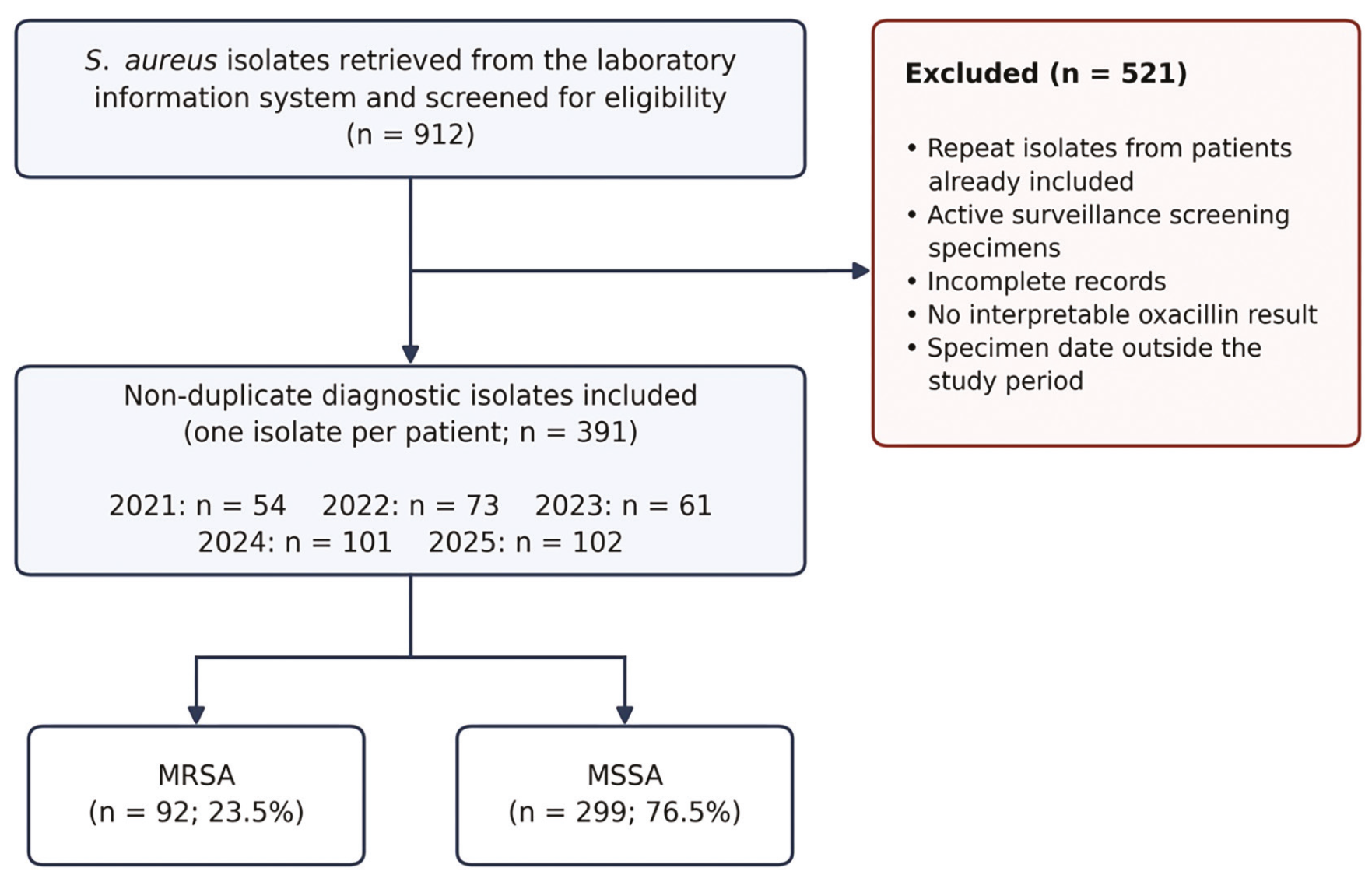
MRSA: Methicillin-resistant S. aureus, MSSA: Methicillin-susceptible S. aureus
Microbiological identification and antimicrobial susceptibility testing
Specimens were cultured on 5% sheep blood agar and eosin methylene blue agar and incubated aerobically at 35-37 °C for 18-24 hours. S. aureus was identified by colony morphology, Gram staining, and catalase and coagulase tests, supplemented with conventional tests when necessary (, ). Species-level identification and antimicrobial susceptibility testing were performed using the BD Phoenix™ automated microbiology system (Becton Dickinson, Sparks, MD, USA) according to the manufacturer’s instructions, and results were interpreted according to European Committee on Antimicrobial Susceptibility Testing (EUCAST) clinical breakpoints (). For longitudinal consistency, categorical results were cross-verified against EUCAST version 15.0 (2025) for agents whose breakpoints did not change during the study period (). Methicillin resistance was determined with the system’s cefoxitin-based screening and oxacillin algorithms. S. aureus ATCC 25923 and ATCC 29213 were used for quality control. All results were reported as susceptible or resistant; no isolate was categorized as “susceptible, increased exposure”. Results for all twelve antimicrobial agents on the panel (erythromycin, clindamycin, ciprofloxacin, levofloxacin, fusidic acid, tetracycline, TMP-SMX, gentamicin, teicoplanin, vancomycin, linezolid and rifampicin) were available for every isolate, and penicillin results were available for 345 isolates (88.2%).
Resistance phenotypes and multiple antibiotic resistance index
MDR, extensive drug resistance (XDR) and pandrug resistance (PDR) were defined at the level of antimicrobial categories according to Magiorakos et al. (). Because methicillin resistance confers resistance to the entire beta-lactam category, MRSA isolates were classified as MDR only when they were also resistant to at least one agent in each of at least two non-beta-lactam antimicrobial categories. The panel used in this study represented eleven antimicrobial categories (anti-staphylococcal beta-lactams, aminoglycosides, ansamycins, fluoroquinolones, folate pathway inhibitors, fusidanes, glycopeptides, lincosamides, macrolides, oxazolidinones and tetracyclines). In contrast, the MAR index was calculated at the level of individual antimicrobial agents, as the number of antimicrobial agents to which the isolate was resistant divided by the total number of antimicrobial agents tested for that isolate (). Oxacillin was excluded to avoid counting the beta-lactam category twice; the denominator was therefore 13 antimicrobial agents (the 12 agents listed above plus penicillin), or 12 agents when penicillin was not tested. Because antimicrobial consumption data were not available, the MAR index and the conventional 0.2 threshold () were used only as descriptive measures.
Ethical considerations
The study protocol was approved by the Non-Drug and Non-Medical Device Research Ethics Committee of Karatay University, acting as the Institutional Review Board (approval date: 26.03.2026; approval number: 2026/037). The study was conducted in accordance with the ethical principles of the Declaration of Helsinki (1975, as revised in 2013). Because the study was retrospective and based on anonymized laboratory records, the ethics committee granted a waiver of written informed consent.
Statistical Analysis
Analyses were performed with IBM SPSS Statistics for Windows, version 22.0 (IBM Corp., Armonk, NY, USA). Categorical variables are presented as number (percentage), and the MAR index as mean ± standard deviation and median [interquartile range (IQR)]; normality was assessed with the Shapiro-Wilk test. Categorical variables were compared with Pearson’s chi-squared test (with Yates’ continuity correction for 2×2 tables) or Fisher’s exact test, and the MAR index with the Mann-Whitney U or Kruskal-Wallis test. Temporal trends were assessed with the chi-squared test for trend (Cochran-Armitage) (). Multivariable binary logistic regression was used to identify factors independently associated with methicillin resistance (age group, sex, specimen type, clinical unit, admission status and calendar year) and to estimate the association between calendar year and resistance to each antimicrobial agent after adjustment for methicillin resistance, admission status and clinical unit; results are presented as adjusted odds ratios (aORs) with 95% confidence intervals (CIs). Pairwise co-resistance among eight antimicrobial agents (28 agent pairs within each of the MRSA and MSSA groups) was quantified with the odds ratio and the phi (Φ) coefficient, tested with Fisher’s exact test and displayed as heatmaps. Because these multiple comparisons were not adjusted for multiplicity, the co-resistance analysis, like the subgroup analyses, was considered exploratory and hypothesis-generating. A two-sided p<0.05 was considered statistically significant. Secondary analyses are described in .
RESULTS
Study population
Of the 912 S. aureus isolates screened, 391 non-duplicate isolates (one per patient) met the inclusion criteria (). The median age of the patients was 51.9 years (IQR 28.3-70.9), and 196 (50.1%) were female. The annual number of isolates increased from 54 in 2021 to 102 in 2025 ().
Table 1. Characteristics of the Staphylococcus aureus isolates and distribution of MRSA and MSSA isolates
Table.1A. All isolates according to year of isolation, age group and clinical unit | - | - | ||
|---|---|---|---|---|
Characteristic | - | - | p-value | p-value |
Year of isolationa | - | - | - | |
2021 | - | - | - | - |
2022 | - | - | - | - |
2023 | - | - | - | - |
2024 | - | - | - | - |
2025 | - | - | - | - |
Age group (years) | - | 0.674 | 0.674 | |
0-18 | - | - | - | - |
19-65 | - | - | - | - |
>65 | - | - | - | - |
Clinical unitb | - | 0.172 | 0.172 | |
Internal medicine units | - | - | - | - |
Surgical units | - | - | - | - |
Intensive care units | - | - | - | - |
Emergency department | - | - | - | - |
Paediatrics | - | - | - | - |
All isolates | - | - | - | - |
Table.1B. MRSA and MSSA isolates according to sex and specimen type | ||||
Characteristic | Total, n (%) | MRSA, n (%) | MSSA, n (%) | p-value |
Sex | 0.700 | |||
Male | 195 (49.9) | 48 (24.6) | 147 (75.4) | - |
Female | 196 (50.1) | 44 (22.4) | 152 (77.6) | - |
Specimen type | 0.709c | |||
Wound | 116 (29.7) | 33 (28.4) | 83 (71.6) | - |
Urine | 103 (26.3) | 23 (22.3) | 80 (77.7) | - |
Blood | 70 (17.9) | 15 (21.4) | 55 (78.6) | - |
Nasald | 37 (9.5) | 5 (13.5) | 32 (86.5) | - |
Deep tracheal aspirate | 28 (7.2) | 7 (25.0) | 21 (75.0) | - |
Sputum | 16 (4.1) | 4 (25.0) | 12 (75.0) | - |
Other specimense | 21 (5.4) | 5 (23.8) | 16 (76.2) | - |
All isolates | 391 (100.0) | 92 (23.5) | 299 (76.5) | - |
Isolate and total columns show column percentages; the MRSA and MSSA columns show row percentages. P-values compare the proportion of MRSA across categories (Pearson’s chi-squared test, with Yates’ continuity correction for 2×2 comparisons). The proportion of MRSA also did not differ between inpatient and outpatient isolates (p=0.961). MRSA: Methicillin-resistant Staphylococcus aureus; MSSA: Methicillin-susceptible Staphylococcus aureus
a: For the association with methicillin resistance, see Table 3. b: Compared across the contributing clinical departments; the infectious diseases department (n=82) is included within internal medicine units. c: Calculated across all 14 specimen categories (Supplementary Table S4). d: Submitted for diagnostic evaluation, not for colonization screening. e: Eight categories with fewer than ten isolates each (joint fluid, bronchial lavage, vaginal, central venous catheter, catheter, aspirate, abscess and pleural fluid cultures)
Distribution of MRSA and MSSA isolates
Overall, 92 isolates (23.5%) were MRSA and 299 (76.5%) were MSSA. Most isolates were recovered from wound (29.7%), urine (26.3%), and blood (17.9%) cultures. The proportion of MRSA did not differ significantly according to age group, sex, admission status, clinical unit, or specimen type ().
Antimicrobial susceptibility testing
Compared with MSSA isolates, MRSA isolates were significantly more resistant to erythromycin, fluoroquinolones, fusidic acid, tetracycline, TMP-SMX, and gentamicin (all p<0.001), whereas clindamycin resistance did not differ significantly (22.8% vs. 16.4%; p=0.210) (). The largest differences were observed for ciprofloxacin (47.8% vs. 3.0%) and TMP-SMX (21.7% vs. 0.7%). No isolate was resistant to vancomycin, linezolid, or rifampicin. Teicoplanin resistance was detected in three isolates (0.8%), which were not available for confirmatory testing.
Table 2. Antimicrobial resistance of MRSA and MSSA isolates [resistant isolates, n (%)]
Antimicrobial agent | All isolates (n=391) | MRSA (n=92) | MSSA (n=299) | p-value |
|---|---|---|---|---|
Erythromycin | 89 (22.8) | 38 (41.3) | 51 (17.1) | <0.001 |
Clindamycin | 70 (17.9) | 21 (22.8) | 49 (16.4) | 0.210 |
Ciprofloxacin | 53 (13.6) | 44 (47.8) | 9 (3.0) | <0.001 |
Levofloxacin | 36 (9.2) | 30 (32.6) | 6 (2.0) | <0.001 |
Fusidic acid | 40 (10.2) | 30 (32.6) | 10 (3.3) | <0.001 |
Tetracycline | 38 (9.7) | 23 (25.0) | 15 (5.0) | <0.001 |
TMP-SMX | 22 (5.6) | 20 (21.7) | 2 (0.7) | <0.001 |
Gentamicin | 21 (5.4) | 13 (14.1) | 8 (2.7) | <0.001 |
Teicoplanin | 3 (0.8) | 2 (2.2) | 1 (0.3) | 0.139 |
Vancomycin | 0 (0.0) | 0 (0.0) | 0 (0.0) | - |
Linezolid | 0 (0.0) | 0 (0.0) | 0 (0.0) | - |
Rifampicin | 0 (0.0) | 0 (0.0) | 0 (0.0) | - |
All remaining isolates were susceptible; no isolate was categorized as susceptible, increased exposure. P-values were obtained with Pearson’s chi-squared test with Yates’ continuity correction or with Fisher’s exact test (teicoplanin). MRSA: Methicillin-resistant Staphylococcus aureus, MSSA: Methicillin-susceptible Staphylococcus aureus, TMP-SMX: Trimethoprim-sulfamethoxazole
Temporal trends in antimicrobial resistance
Resistance to ciprofloxacin, erythromycin, clindamycin and fusidic acid increased significantly between 2021 and 2025 (p for trend <0.05), with the largest increase observed for ciprofloxacin (from 7.4% to 26.5%; p<0.001) (). No significant trend was found for levofloxacin, tetracycline, gentamicin, or TMP-SMX. Annual resistance rates with 95% CIs are provided in .
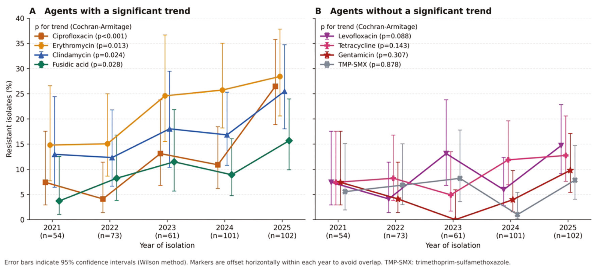
TMP-SMX: Trimethoprim-sulfamethoxazole, MRSA: Methicillin-resistant Staphylococcus aureus, MSSA: Methicillin-susceptible Staphylococcus aureus
Multivariable analysis
Calendar year was the only factor independently associated with methicillin resistance (aOR 1.27 per year; 95% CI 1.04-1.55; p=0.017) (). After adjustment for methicillin resistance, admission status and clinical unit, calendar year remained independently associated with resistance to ciprofloxacin (aOR 1.60), clindamycin (aOR 1.28) and erythromycin (aOR 1.25), but not with resistance to levofloxacin or TMP-SMX. Methicillin resistance was strongly associated with resistance to non-beta-lactam antimicrobial agents, most markedly TMP-SMX (aOR 55.8) and ciprofloxacin (aOR 37.2).
Table 3. Multivariable logistic regression analysis of factors associated with methicillin resistance and with resistance to individual antimicrobial agents
Outcome and variable | aOR (95% CI) | p-value |
|---|---|---|
Methicillin resistance (MRSA vs. MSSA)a | - | |
Calendar year (per 1-year increase) | 1.27 (1.04-1.55) | 0.017 |
Resistance to individual agents: calendar year (per 1-year increase)b | - | |
Ciprofloxacin | 1.60 (1.16-2.21) | 0.004 |
Clindamycin | 1.28 (1.04-1.57) | 0.018 |
Erythromycin | 1.25 (1.03-1.52) | 0.021 |
Levofloxacin | 1.08 (0.78-1.48) | 0.648 |
TMP-SMX | 0.78 (0.54-1.14) | 0.207 |
Resistance to individual agents: methicillin resistance (MRSA vs. MSSA)b,c | - | |
TMP-SMX | 55.8 (12.1-257.9) | <0.001 |
Ciprofloxacin | 37.2 (15.9-87.2) | <0.001 |
a: Single model including calendar year, age group, sex, specimen type, clinical unit and admission status; age group, sex, specimen type, clinical unit and admission status were not significantly associated with methicillin resistance. b: Separate models for each agent including calendar year and methicillin resistance, adjusted for admission status and clinical unit. c: The two agents with the strongest associations are shown. aOR: Adjusted odds ratio, CI: Confidence interval, MRSA: Methicillin-resistant Staphylococcus aureus, MSSA: Methicillin-susceptible Staphylococcus aureus, TMP-SMX: Trimethoprim-sulfamethoxazole
MAR index and multidrug resistance
The median MAR index was 0.077 (IQR 0.077-0.231), and 99 isolates (25.3%) had a MAR index above 0.2. The MAR index was higher in MRSA than in MSSA isolates (mean 0.261 vs. 0.105; p<0.001) but did not differ significantly between clinical units (p=0.597). MDR was identified in 51 MRSA (55.4%) and 9 MSSA (3.0%) isolates (p<0.001), and no isolate met the XDR or PDR criteria (Supplementary Table S2). Secondary analyses are summarized in Supplementary Results S1.
Co-resistance associations
Pairwise co-resistance associations among the eight antimicrobial agents included in this exploratory analysis are shown in , and detailed statistics are given in . Of the 28 agent pairs examined in each group, 15 in MRSA and 8 in MSSA isolates showed a nominally significant association (unadjusted p<0.05). The strongest associations were between clindamycin and erythromycin (Φ=0.60 in MRSA and 0.90 in MSSA isolates; p<0.001 for both), with more than 93% of clindamycin-resistant isolates in both groups also being resistant to erythromycin, and between ciprofloxacin and levofloxacin (Φ=0.68 and 0.81, respectively; p<0.001 for both). Other associations, such as those between gentamicin and fluoroquinolone resistance (Φ=0.30-0.42) and, in MRSA isolates, between fusidic acid and levofloxacin resistance (Φ=0.51), should be interpreted with caution given the number of comparisons.
Discussion
In this five-year single-center study, MRSA accounted for 23.5% of clinical S. aureus isolates, and its proportion increased over the study period; this association persisted after adjustment for patient and unit characteristics. MRSA isolates carried most of the resistance burden: more than half were MDR, and methicillin resistance was strongly associated with resistance to most non-beta-lactam antimicrobial agents. Resistance to ciprofloxacin, erythromycin and clindamycin increased over time, and these associations persisted after adjustment for methicillin resistance, whereas no resistance to vancomycin, linezolid or rifampicin was detected.
The MRSA proportion observed here lies within the 20-40% range reported for Türkiye and other countries of Southern Europe, the Eastern Mediterranean and North Africa in WHO and European surveillance data, whereas proportions below 10% have been reported in Northern European countries with long-standing stewardship and infection-control programmes (, ). MRSA remains one of the leading pathogen-drug combinations responsible for deaths attributable to AMR worldwide (, ). Whereas MRSA proportions have declined in many European countries and in North America since the mid-2000s (, ), the proportion of MRSA in our cohort increased during the study period. Genomic studies from Saudi Arabia have linked recent increases in MRSA to clonal expansion and plasmid-mediated dissemination of resistance (, ); in the absence of molecular typing, we cannot determine whether a similar process, changes in referral patterns or an increase in community-associated MRSA underlies our observation (, ).
As reported previously, MRSA isolates were markedly more resistant than MSSA isolates to non-beta-lactam antimicrobial agents (, ). Gentamicin resistance was approximately fivefold higher in MRSA; similar differences have been attributed to the co-location of aminoglycoside-modifying enzyme genes with other resistance determinants on mobile genetic elements (, ), although resistance genes were not investigated in the present study. TMP-SMX, which is among the oral agents used for MRSA skin and soft tissue infections (), showed resistance in about one in five MRSA isolates (21.7%); because the TMP-SMX breakpoint was revised after the study period, this rate should be compared with other reports with caution. The increase in ciprofloxacin resistance, which persisted after adjustment for methicillin resistance, is in line with the increasing fluoroquinolone resistance reported among S. aureus isolates in other settings (). Almost all levofloxacin-resistant isolates were also resistant to ciprofloxacin, a pattern that may reflect class-wide target-site mutations in gyrA and grlA (the parC homologue) (); however, this could not be confirmed without molecular analysis.
More than 93% of clindamycin-resistant isolates in both the MRSA and MSSA groups were also resistant to erythromycin, a pattern compatible with macrolide-lincosamide-streptogramin B resistance mediated by erm genes (, ); because erm genes were not investigated, this interpretation remains presumptive. In addition, inducible clindamycin resistance was not analysed as a separate phenotype, so its contribution to the observed resistance rates could not be assessed. The increase in fusidic acid resistance, which was associated with levofloxacin resistance in MRSA isolates in the exploratory co-resistance analysis, has been attributed elsewhere to selective pressure from widespread topical use () and to co-selection within multiresistant strains; because antimicrobial consumption and molecular data were not available, neither explanation could be evaluated in this study. No vancomycin-resistant isolate was detected; however, reduced vancomycin susceptibility, which has been investigated in MRSA isolates from Türkiye (), cannot be excluded by routine automated testing.
The MAR index showed that the accumulated resistance burden was concentrated in MRSA rather than in particular clinical units. Because antimicrobial consumption data were not available, the 0.2 threshold, originally proposed to identify high-risk sources of contamination (), was used only descriptively, as in recent clinical applications of the index (). Likewise, MRSA proportions were similar across age groups, sexes, specimen types and clinical units, a finding compatible with reports of MRSA outside traditional high-risk groups, including paediatric skin and soft tissue infections in Türkiye (), and with international data (); however, without clinical and molecular data, the community or healthcare origin of these isolates could not be determined.
These findings describe the in vitro susceptibility of isolates recovered from diagnostic specimens at a single center. Because clinical infection criteria, antimicrobial exposure data, and molecular typing were not available, they do not permit conclusions about treatment effectiveness or about the causes of the observed temporal changes.
Strengths
The main strengths of this study are as follows:
• A five-year dataset restricted to the first isolate per patient, with susceptibility testing on a single automated platform and harmonization of categorical results to one EUCAST version.
• Multivariable adjustment of temporal trends for methicillin resistance, admission status, and clinical unit.
• Combined use of the category-based MDR classification, the agent-based MAR index and exploratory co-resistance mapping to describe the resistance burden.
Study Limitations
The main limitations are as follows:
• Retrospective, single-center design in a tertiary referral hospital, which may limit generalizability and introduce referral bias.
• Lack of clinical data (clinical infection criteria, comorbidities, prior antimicrobial use, and outcomes) and antimicrobial consumption data; colonization could therefore not be distinguished from infection, and resistance could not be linked to antimicrobial exposure.
• No molecular confirmation of methicillin resistance (mecA/mecC) or genotyping; clonal relatedness and resistance mechanisms (e.g., erm genes or fluoroquinolone target-site mutations) could not be assessed.
• Unequal annual sample sizes, which may have affected trend estimates.
• Multiple pairwise comparisons in the co-resistance analysis without formal adjustment for multiplicity; these associations are exploratory, and some nominally significant findings may have arisen by chance.
• Minimum inhibitory concentrations were not archived for most agents and isolates were not stored; results could not be re-interpreted against later breakpoint revisions (e.g., TMP-SMX in EUCAST v16.0), and the three teicoplanin-resistant isolates could not be confirmed.
Conclusion
In this center, MRSA accounted for about one quarter of clinical S. aureus isolates, and its proportion increased over the five-year study period. Methicillin resistance was associated with higher rates of resistance to most non-beta-lactam antimicrobial agents and with multidrug resistance. Resistance to ciprofloxacin, erythromycin and clindamycin increased over time and remained associated with calendar year after adjustment for methicillin resistance, whereas no resistance to vancomycin, linezolid or rifampicin was detected.
Footnotes
Ethics
Ethics Committee Approval: The study protocol was approved by the Non-Drug and Non-Medical Device Research Ethics Committee of Karatay University, acting as the Institutional Review Board (approval date: 26.03.2026; approval number: 2026/037). The study was conducted in accordance with the ethical principles of the Declaration of Helsinki (1975, as revised in 2013).
Informed Consent: Because the study was retrospective and based on anonymized laboratory records, the ethics committee granted a waiver of written informed consent.
Authorship Contributions
Surgical and Medical Practices: B.E., Concept: B.E., R.B., Design: B.E., R.B., Data Collection or Processing: B.E., Analysis or Interpretation: B.E., R.B., Literature Search: B.E., R.B., Writing: B.E.
Conflict of Interest: No conflict of interest was declared by the authors.
Financial Disclosure: The authors declared that this study received no financial support.
References
World Health Organization. Global antimicrobial resistance and use surveillance system (GLASS) report 2023 [Internet]. Geneva: World Health Organization; 2023 [cited 2026 Mar 15]. Available from: https://www.who.int/initiatives/glass
Antimicrobial Resistance Collaborators. Global burden of bacterial antimicrobial resistance in 2019: a systematic analysis. Lancet. 2022;399(10325):629-655. Erratum in: Lancet. 2022;400(10358):1102.
O’Neill J. Antimicrobial resistance: tackling a crisis for the health and wealth of nations. London: review on antimicrobial resistance; 2014.
World Health Organization. WHO bacterial priority pathogens list 2024: bacterial pathogens of public health importance to guide research, development and strategies to prevent and control antimicrobial resistance [Internet]. Geneva: World Health Organization; 2024 [cited 2026 Mar 15]. Available from: https://www.who.int/publications/i/item/9789240093461
Oyenuga N, Cobo-Díaz JF, Alvarez-Ordóñez A, Alexa EA. Overview of antimicrobial resistant ESKAPEE pathogens in food sources and their implications from a one health perspective. Microorganisms. 2024;12(10):2084.
Tunçer G, Geyiktepe Güçlü C, Sürme S, Sayılı U, Yücel Ç, Güçlü KG, et al. The more vulnerable, the more fatal: predictors for mortality in patients with dreadful infective endocarditis. FLORA. 2024;29(3):380-387.
Russo TA, Broczkowski B, Sallerson S, Knee A, Lorenzo MP. Association between erythromycin or clindamycin resistance and 30-day mortality in patients with MSSA bacteremia. J Pharm Pract. 2026;39(1):18-24.
Sancak B. Staphylococcus aureus and antibiotic resistance. Mikrobiyol Bul. 2011;45(3):565-576.
Jevons MP. “Celbenin”-resistant staphylococci. Br Med J. 1961;1(5219):124-125.
Alhejaili AY, Zhou G, Halawa H, Huang J, Fallatah O, Hirayban R, et al. Methicillin-resistant Staphylococcus aureus in Saudi Arabia: genomic evidence of recent clonal expansion and plasmid-driven resistance dissemination. Front Microbiol. 2025;16:1602985.
Foster TJ. Antibiotic resistance in Staphylococcus aureus: current status and future prospects. FEMS Microbiol Rev. 2017;41(3):430-449.
David MZ, Daum RS. Community-associated methicillin-resistant Staphylococcus aureus: epidemiology and clinical consequences of an emerging epidemic. Clin Microbiol Rev. 2010;23(3):616-687.
Carrel M, Smith M, Shi Q, Hasegawa S, Clore GS, Perencevich EN, et al. Antimicrobial resistance patterns of outpatient Staphylococcus aureus isolates. JAMA Netw Open. 2024;7(6):e2417199.
Krumperman PH. Multiple antibiotic resistance indexing of Escherichia coli to identify high-risk sources of fecal contamination of foods. Appl Environ Microbiol. 1983;46(1):165-170.
von Elm E, Altman DG, Egger M, Pocock SJ, Gøtzsche PC, Vandenbroucke JP; STROBE Initiative. The strengthening the reporting of observational studies in epidemiology (STROBE) statement: guidelines for reporting observational studies. Lancet. 2007;370(9596):1453-1457.
Clinical and Laboratory Standards Institute. Analysis and presentation of cumulative antimicrobial susceptibility test data. 5th ed. CLSI guideline M39. Wayne (PA): Clinical and Laboratory Standards Institute; 2022.
Bahçeci İ, Aksoy D, Karaca Semerci E, Yıldız S, Alpdoğan YE, Duran ÖF, et al. Antimicrobial resistance in Staphylococcus aureus strains isolated from blood cultures: a three-year study. Rize Medical Journal. 2023;1(4):6-19.
Procop GW, Church DL, Hall GS, Janda WM, Koneman EW, Schreckenberger PC, et al. Koneman’s color atlas and textbook of diagnostic microbiology. 7th ed. Philadelphia: Wolters Kluwer; 2017.
The European Committee on Antimicrobial Susceptibility Testing. Breakpoint tables for interpretation of MICs and zone diameters. Version 15.0 [Internet]. EUCAST; 2025 [cited 2026 Sep 18]. Available from: https://www.eucast.org/clinical_breakpoints
Magiorakos AP, Srinivasan A, Carey RB, Carmeli Y, Falagas ME, Giske CG, et al. Multidrug-resistant, extensively drug-resistant and pandrug-resistant bacteria: an international expert proposal for interim standard definitions for acquired resistance. Clin Microbiol Infect. 2012;18(3):268-281.
Armitage P. Tests for linear trends in proportions and frequencies. Biometrics. 1955;11(3):375-386.
European Centre for Disease Prevention and Control. Antimicrobial resistance surveillance in Europe 2023: 2021 data [Internet]. Stockholm: ECDC; 2022 [cited 2026 Mar 15]. Available from: https://www.ecdc.europa.eu/en/publications-data/antimicrobial-resistance-surveillance-europe-2023-2021-data
GBD 2021 Antimicrobial Resistance Collaborators. Global burden of bacterial antimicrobial resistance 1990-2021: a systematic analysis with forecasts to 2050. Lancet. 2024;404(10459):1199-1226.
Diekema DJ, Pfaller MA, Shortridge D, Zervos M, Jones RN. Twenty-year trends in antimicrobial susceptibilities among Staphylococcus aureus from the SENTRY antimicrobial surveillance program. Open Forum Infect Dis. 2019;6(Suppl 1):S47-S53. Erratum in: Open Forum Infect Dis. 2019;6(5):ofz202. Zervos, Marcos [corrected to Zervos, Marcus].
Alkuraythi DM, Alkhulaifi MM, Altwiley DA, Alarwi M, Mujallad MI, Alshomrani MK, et al. Genomic and epidemiological insights into the emergence and dominance of MRSA clones in Riyadh’s healthcare facilities. Sci Rep. 2026;16(1):3835.
Li J, Cheng F, Wei X, Bai Y, Wang Q, Li B, et al. Methicillin-resistant Staphylococcus aureus (MRSA): resistance, prevalence, and coping strategies. Antibiotics. 2025;14(8):771.
Ramirez MS, Tolmasky ME. Aminoglycoside modifying enzymes. Drug Resist Updat. 2010;13(6):151-171.
Tong SY, Davis JS, Eichenberger E, Holland TL, Fowler VG Jr. Staphylococcus aureus infections: epidemiology, pathophysiology, clinical manifestations, and management. Clin Microbiol Rev. 2015;28(3):603-661.
Hooper DC, Jacoby GA. Mechanisms of drug resistance: quinolone resistance. Ann N Y Acad Sci. 2015;1354(1):12-31.
Khursheed M, Sulaiman M, Alhassan AM, Al Awad TH, Ibrahim AE, Wilson G, et al. Clindamycin-resistant among Staphylococcus aureus: investigation into phenotypic and genotypic profiles. PLoS One. 2025;20(8):e0329467.
Leclercq R. Mechanisms of resistance to macrolides and lincosamides: nature of the resistance elements and their clinical implications. Clin Infect Dis. 2002;34(4):482-492.
Howden BP, Grayson ML. Dumb and dumber--the potential waste of a useful antistaphylococcal agent: emerging fusidic acid resistance in Staphylococcus aureus. Clin Infect Dis. 2006;42(3):394-400.
Çıkman A, Aydın M, Gülhan B, Parlak M, Gültepe B, Kalaycı Y, et al. Metisiline dirençli Staphylococcus aureus izolatlarının antibiyotik direnci ve azalmış vankomisin duyarlılığının araştırılması: çok merkezli bir çalışma [Investigation of antibiotic resistance patterns and reduced vancomycin susceptibilities of methicillin-resistant Staphylococcus aureus isolates: a multi-center study]. Mikrobiyol Bul. 2015;49(2):240-248.
Isaiah DO, Otokunefor K, Agbagwa OE. Multiple antibiotic resistance indexing and molecular identification of Escherichia coli isolated from clinical and nonclinical sources in Port Harcourt Metropolis, Nigeria. Pan Afr Med J. 2025;51:11.
Yakut N, Ergenç Z, Bayraktar S, Akbolat İ, Sayın E, İlki A, et al. Antimicrobial susceptibility and characterization of skin and soft tissue infections caused by Staphylococcus aureus in children. FLORA. 2024;29(1):85-95.
Supplementary Materials
Supplementary Table S1
Download