
Abstract
Aims
In forensic investigations, ABO blood grouping from biological evidence plays an important role. The aim of this study is to evaluate the feasibility of ABO blood group determination from tear samples and to assess the effect of different environmental conditions on the detectability of ABO antigens.
Methods
This is a laboratory-based experimental cross-sectional study. Participants were healthy individuals aged between 18 and 50 years with a known ABO blood group who gave consent for the study and were included in the study. Non-secretor individuals (identified by analyzing saliva samples) or individuals with ocular or oral disorders, have recent eye surgeries and using any eye drops and other eye medications, individuals using oral medication, which can affect the saliva composition were excluded from the study. Tear samples were collected on sterile tissue paper, and each sample was divided into five portions. Samples maintained at room temperature were analyzed immediately (0 hours) and served as the baseline control, while the remaining portions were exposed to low temperature (4 °C), high temperature (56 °C), dry, or moist conditions for 24 hours. ABO blood grouping was performed using the absorption-elution technique.
Results
A total of 66 participants were included in the study (mean age: 24.2±6.9 years; 51 females [77.3%]). At room temperature the ABO antigens were detected in 57 samples of 66 (86.4%), at low and high temperature it was observed in 55 and 52 of 66 samples (83.3% and 78.8%), in dry and moist condition it was observed in 53 and 43 of 66 samples (80.3% and 65.2%). Cochran’s Q test demonstrated a significant overall difference among conditions (Q=11.72, df=4, p=0.0196).
Conclusions
The results show that ABO blood group antigens can be successfully detected in tear samples under controlled conditions. However, the reduction in detectability, particularly under moist conditions, highlights the influence of the environment.
Keywords: Tear stains, ABO antigens detection, absorption-elution technique, environmental factors, secretor status, forensic serology
Introduction
Biological fluids, such as blood, saliva, and semen, play a crucial role in forensic investigations by helping in individual identification and crime scene reconstruction. Among the several serological markers, the ABO blood group system remains one of the most widely used tools in forensic science because of its simplicity, cost-effectiveness and reliability (-). Although DNA profiling has become the gold standard for individual identification, serological techniques such as ABO blood typing continue to provide important preliminary information and help to narrow down the suspect pool in forensic cases, particularly in scenarios when DNA analysis is not possible due to sample degradation, limited sample quantity or laboratory limitations ().
At crime scenes, recovered biological evidence is not always blood; alternative body fluids, such as saliva, semen, sweat, and urine, may also be encountered. Many of these body fluids contain soluble ABO antigens in individuals with secretor status (). Studies show that approximately 80% of the population are secretors, meaning these individuals express ABO glycoproteins in body secretions due to the functional activity of the FUT2 gene (, ). This gene activity enables ABO blood grouping from non-blood body fluids in the secretor population.
Among the various non-blood biological fluids, tear fluid has recently gained attention as a potential forensic sample. Tears are usually shed in circumstances such as emotional stress and physical trauma, which are common occurrences during criminal activities (, ). Tears contain water, proteins, glycoproteins, lipids, and electrolytes. They share biochemical similarities with other body fluids, which are often used in forensic investigations (, ). Previous studies have established the detection, spectral analysis, and DNA profiling of tear stains, highlighting their forensic relevance ().
Despite these developments, the application of tear fluid in serological tests remains underexplored. No studies have reported the presence of ABO antigens in tears, and no information is known about their stability under environmental conditions (). Biological samples recovered from crime scenes are typically subjected to environmental factors such as temperature fluctuations, humidity, sunlight exposure, and microbial activity, which may affect antigen stability and detection.
Since tear fluid contains soluble antigens in relatively low concentrations compared to blood (), they may be more vulnerable to environmental degradation. It is therefore essential to understand the effects of environmental conditions on the stability and detectability of tear-based ABO antigens to determine their forensic reliability.
Hence, the present study aims to evaluate the feasibility of ABO blood grouping from tear samples exposed to environmental conditions such as temperature and moisture using the absorption-elution (AE) technique, and to determine how these factors influence antigen detectability.
Methods
Study design and setting
This is a laboratory-based experimental cross-sectional study conducted in the department of forensic medicine and toxicology from 25.9.2025 to 25.2.2026.
Ethical approval
Ethical approval for the study was obtained from the Institutional Ethics Committee of Kasturba Medical College (IEC no: IEC2-446, date: 23.09.2025). Written informed consent was obtained from all participants prior to sample collection. The study protocol conforms to the ethical guidelines of the 1975 Declaration of Helsinki (Helsinki Declaration as revised in 2013). Informed consent to participate was written and was given to each participant.
Study population and eligibility criteria
The study population consisted of 66 healthy volunteers from the student and staff population of Kasturba Medical College, Manipal, aged between 18-50 years with known ABO blood groups. Healthy individuals aged between 18-50 years, with known ABO blood group and those who gave informed consent were included in the study. Non-secretor individuals (identified by analyzing saliva samples), individuals with ocular or oral disorders, having history of recent eye surgeries or participants using eye drops and other eye medications, using oral medication, which can affect the saliva composition, and who have consumed food or beverages (except water) 30 minutes before saliva collection were excluded from the study.
Sample collection
Unstimulated saliva samples were first collected in sterile test tubes for determining secretor status. Following this, tear samples were collected onto sterile two-ply tissue paper, and the stained area was marked for subsequent analysis. Participants were instructed to gently close their eyes and perform eye movements to stimulate tear flow. When necessary, mild menthol balm was applied externally, without direct contact with the eyes, to induce reflex tearing while ensuring that tear composition remained unaltered.
Environmental exposure conditions
Each tear sample was cut and divided into five experimental groups. Samples maintained at room temperature served as the baseline control and were analyzed immediately (0 hours). The remaining samples were subjected to different environmental conditions for 24 hours:
1. High temperature: Tissue fragments were incubated at 56 °C in a hot air oven.
2. Low temperature: Samples were stored at 4 °C.
3. Dry condition: Tissue fragments were wrapped in brown paper to permit airflow and stored at room temperature.
4. Moist condition: Tissue fragments were placed in a humid chamber (Petri dish containing moistened filter paper) and stored at 4-8 °C.
Secretor status determination
Secretor status was determined using the standard absorption-inhibition technique. Saliva samples were heat-inactivated at 56 °C and centrifuged at 3000 rpm for 2-3 minutes. The saliva extract was incubated with anti-H antisera, followed by the addition of O-group indicator red blood cells. Absence of agglutination was interpreted as a positive secretor status.
For quality control, known secretor and non-secretor saliva samples were included as positive and negative controls, respectively. All agglutination reactions were assessed independently by two trained observers. In cases of disagreement, the test was repeated and a consensus interpretation was reached.
ABO blood grouping from tear samples
ABO blood grouping of tear samples was performed using the AE technique. Following environmental exposure, each tissue fragment containing tear stains was incubated overnight at 4 °C with anti-A, anti-B, and anti-H antisera in separate test tubes. The samples were subsequently washed with chilled saline solution to remove unbound antibodies. Bound antibodies were eluted at 56 °C in a water bath for 15-20 minutes and tested against appropriate indicator red cell suspensions. Agglutination reactions were examined both macroscopically () and microscopically.
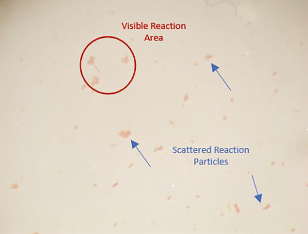
To ensure reliability of the results, known blood group samples served as positive controls, while unstained tissue fragments processed under identical conditions served as negative controls. Agglutination reactions were graded according to standard serological criteria by two trained observers who were blinded to the secretor status and environmental exposure condition of the samples. All tests were performed in duplicate, and discrepant results were re-evaluated through repeat testing to confirm reproducibility.
Statistical Analysis
The data was recorded in Microsoft Excel and analyzed using Jamovi software (version 2.4). Detection frequencies and percentages were calculated to compare antigen detectability across different environmental conditions. Cochran’s Q test was used to demonstrate variations among different environmental conditions. Pairwise McNemar comparisons of different environmental conditions with Bonferroni correction were also done.
Results
Demographic details of study participants
A total of 66 participants (15 males, 51 females) were included in the study. The mean age of the participants was 24.2±6.9 years.
Tear samples were collected from each participant and were exposed to five environmental conditions: room temperature, low temperature (4 °C), high temperature (56 °C), dry condition and moist condition. All the samples were analyzed for ABO blood group detection using the AE technique.
The distribution of ABO blood groups among participants showed that blood group O was the most common (37.9%), followed by A (30.3%) and B (28.8%), while AB was the least represented (3.0%).
Detection of ABO antigens in tear samples
ABO blood group antigens were detected in tear samples across all blood groups under at least one testing condition, indicating the presence of soluble blood group substances in tear fluid. At room temperature, agglutination was observed in 57 of 66 samples (86.4%), indicating a high rate of antigen detectability. These results suggest that tear fluid contains sufficient soluble ABO antigens to test blood grouping using the AE method.
Detection of ABO antigens under different environmental conditions
Environmental exposure has influenced the detectability of ABO antigens in tear samples. At room temperature, the ABO antigens were detected in 57 of 66 samples (86.4%), at low temperature in 55 of 66 samples (83.3%), and at high temperature in 52 of 66 samples (78.8%). In dry and moist conditions, ABO antigen detection was observed in 53 of 66 samples (80.3%) and 43 of 66 samples (65.2%).
When compared together (), the results showed that environmental conditions significantly influenced agglutination outcomes. Cochran’s Q test demonstrated a significant overall difference among conditions (Q=11.72, df=4, p=0.0196). Moist conditions demonstrated the lowest positivity rates. Room-temperature and low-temperature conditions maintained higher positivity rates. Pairwise comparisons between environmental conditions using McNemar’s test with Bonferroni correction are presented in . From the table, it is evident that only a statistically significant pairwise comparison after correction was observed for room temperature and moist condition (p=0.0258).
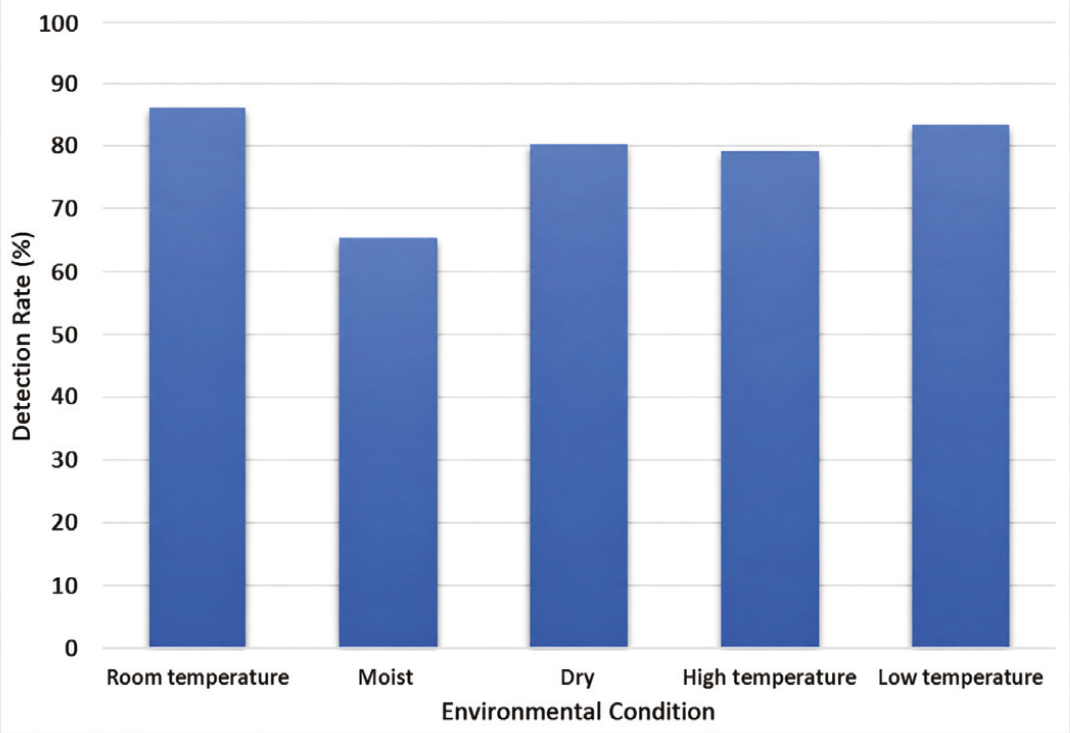
Table 1. Pairwise comparisons of ABO antigen detection across environmental conditions using McNemar’s test with Bonferroni correction
Condition A* | Condition B* | A-/B+* | A+/B−* | Unadjusted p-value | Bonferroni-adjusted p-value |
|---|---|---|---|---|---|
RT | M | 3 | 17 | 0.0026 | 0.0258 |
RT | D | 8 | 12 | 0.5034 | 1.0000 |
RT | HT | 7 | 12 | 0.3593 | 1.0000 |
RT | LT | 8 | 10 | 0.8145 | 1.0000 |
M | D | 16 | 6 | 0.0525 | 0.5248 |
M | HT | 13 | 4 | 0.0490 | 0.4904 |
M | LT | 18 | 6 | 0.0227 | 0.2266 |
D | HT | 10 | 11 | 1.0000 | 1.0000 |
D | LT | 9 | 7 | 0.8036 | 1.0000 |
HT | LT | 12 | 9 | 0.6636 | 1.0000 |
*: A and B refer to the compared environmental conditions, and “+” and “-” indicate positive and negative detection results. RT: Room temperature, HT: High temperature, LT: Low temperature, D: Dry condition, M: Moist condition
Positivity rates in different environmental conditions of various blood groups are shown in . AB+ samples showed 100% positivity under all conditions. B- samples also maintained nearly 100% positivity under RT, moist, dry, and high-temperature conditions; however, positivity declined markedly to 50% under low-temperature conditions. B+ samples demonstrated 100% positivity at RT, followed by lower values under dry and high-temperature conditions (approximately 94%), moist conditions (approximately 89%), and low-temperature conditions (approximately 88%). A+ samples showed the highest positivity under low-temperature conditions (90%), followed by RT (85%), dry and high-temperature conditions (80% each), and moist conditions (75%). O+ samples exhibited (approximately 75%) positivity at RT and low temperature, followed by dry (approximately 68%) and high-temperature conditions (approximately 64%), while moist conditions produced the lowest positivity (approximately 36%).
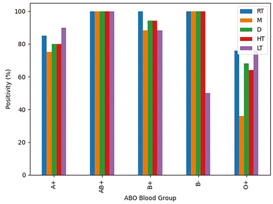
RT: Room temperature, M: Moist condition, D: Dry condition, HT: High temperature, LT: Low temperature
Discussion
The findings of the present study demonstrate that ABO blood group antigens can be detected in tear samples using the AE technique, suggesting the potential of tear fluid as an alternative biological sample for ABO blood group identification.
The study demonstrated successful detection of ABO antigens in tear samples from individuals belonging to all ABO blood groups. These findings support the presence of soluble blood group substances in tear fluid and are consistent with the known expression of secretor-related antigens in body secretions (). The presence of these soluble antigens enables specific antigen-antibody interactions, allowing their identification through sensitive serological methods such as the AE technique ().
The AE technique was found to be effective for the analysis of tear samples, which typically contain relatively low concentrations of soluble blood group antigens. Its capacity to detect and recover weakly expressed antigens from limited or partially degraded biological material highlights its value in forensic serology, particularly when only small quantities of evidence are available for examination (, ).
Environmental conditions were found to influence the stability and subsequent detectability of ABO antigens in tear samples. Among the conditions evaluated, samples maintained at room temperature exhibited the highest detection rate (86.4%), indicating that short-term exposure to ambient environmental conditions has minimal impact on antigen preservation (, ). This finding suggests that freshly deposited tear stains may retain sufficient antigenic integrity for ABO blood group determination and could therefore provide valuable forensic information when collected and analyzed within a relatively short period after deposition.
Samples stored under low-temperature conditions (4 °C) exhibited antigen detection rates that were comparable to those observed at room temperature. This finding is consistent with the well-established role of low temperatures in inhibiting enzymatic activity and microbial growth, thereby enhancing the preservation of biological markers over time (, ). Conversely, samples exposed to elevated temperatures (56 °C) demonstrated a moderate reduction in antigen detectability (78.8%). High temperatures can induce structural modifications in glycoproteins and carbohydrate components that contribute to ABO antigen specificity, potentially impairing antigen-antibody binding and reducing the strength of agglutination reactions (, , ). Dry environmental conditions also supported relatively good antigen preservation, likely because the absence of moisture limits microbial proliferation and slows biochemical degradation processes, thereby helping maintain antigen stability ().
Among the environmental conditions evaluated, samples maintained under moist conditions exhibited the lowest antigen detection rate (65.2%). The presence of moisture is known to facilitate enzymatic activity, hydrolytic processes, and microbial growth, all of which can contribute to the accelerated degradation of soluble blood group antigens in biological samples (, ). Consequently, prolonged exposure to humid environments may compromise antigen stability and reduce the likelihood of successful ABO blood group determination from tear stains.
Various studies have been conducted to identify ABO blood grouping from various body tissues. Velani et al. () conducted a study to evaluate the accuracy of ABO blood group determination and Rh typing from dried salivary samples. The study identified that there was a 100% positive correlation for ABO blood grouping, but a mere 14.81% positive correlation for Rh typing between the dried salivary samples and the extracted blood (). Singh and Bhist () studied the accuracy of dried saliva samples in establishing ABO and Rh blood types. The findings revealed that saliva and blood typing for ABO groups corresponded nearly perfectly, highlighting the promise of saliva-based ABO identification, particularly for secretors. However, the correlation between the Rh type from saliva and the blood study was significantly weaker (). Sowndharya and Chawla () conducted a study to check the accuracy of saliva samples in establishing ABO and Rh blood types. The findings revealed that 100% blood grouping can be determined from saliva samples ().
Rao et al. () conducted a study to ascertain the ABO blood group from both the hard (dentin) and soft tissue (pulp) of the tooth by using the AE technique. They found that the dentin and pulp were shown to have positive blood group antigens for the ABO and Rh factors. While neither pulp nor dentin performed significantly differently in identifying the blood group antigens, pulp showed marginally higher accuracy (). Mishra et al. () conducted a study to ascertain ABO blood group from extracted teeth using pulp and dentin tissues with the help of the AE technique. They found that 54 teeth, that is, 90% of the total sample, were positive in the estimation of blood group (). Ramnarayan et al. () conducted a study to detect ABO blood group substances from soft and hard tissues of teeth. They found that pulp showed a very large correlation in both fresh and long-standing teeth. Hard tissue showed a large correlation in both groups, indicating that hard tissue is quite reliable for detecting blood group and that there is not much difference in reliability between the groups. However, combining pulp and hard tissue, correlation was moderate ().
To the best of our knowledge, no previous studies have investigated ABO blood group determination from tear samples. The findings of the present study therefore provide preliminary evidence supporting the forensic applicability of tear fluid as an alternative biological specimen for ABO blood group analysis. Nevertheless, the influence of environmental conditions should be carefully considered when interpreting blood grouping results obtained from tear evidence. Overall, the results suggest that ABO antigens in tear samples exhibit considerable stability under certain environmental conditions, particularly at room and low temperatures. In contrast, exposure to moist conditions was associated with a marked reduction in antigen detectability, indicating that humidity may adversely affect antigen preservation and, consequently, the reliability of ABO blood group determination.
Study Limitations
Despite these findings, several limitations of the present study should be acknowledged. The study focused primarily on the detection of ABO antigens in tear samples and did not evaluate quantitative changes in antigen concentration or the degree of antigen expression under different environmental conditions. In addition, the sample size was relatively small, which limited the statistical power of the study and prevented the application of inferential statistical analyses to assess associations between variables. The composition of the study sample also limits the generalizability of the findings. Only secretor individuals were included in the study; therefore, the results cannot be extrapolated to non-secretor populations, who may exhibit different patterns of antigen detectability in tear samples. Furthermore, the number of participants belonging to the AB blood group was very limited, reducing the ability to draw robust conclusions regarding the persistence and detectability of AB antigens under the tested conditions. Consequently, the findings should be interpreted with caution, particularly when considering their applicability to broader and more diverse populations. The environmental simulation model employed in this study was designed to represent selected conditions that may be encountered during forensic investigations, including variations in temperature and moisture. However, samples were exposed to controlled laboratory conditions for only 24 hours, which may not adequately reflect the complexity of actual crime scene environments. Real-world forensic samples are often subjected to multiple environmental influences simultaneously, including sunlight and ultraviolet radiation, fluctuations in temperature and humidity, different substrate materials, microbial activity, and prolonged periods of exposure. The present study evaluated only a limited number of environmental variables independently and did not assess the combined effects of these factors. Therefore, the observed results may not fully represent antigen stability under real forensic conditions. Future studies involving larger and more balanced sample populations, inclusion of non-secretors, longer exposure periods, diverse substrates, and more comprehensive environmental simulations are warranted to validate and extend the current findings.
Conclusion
This study demonstrated that tear fluid may serve as an alternative biological sample for ABO blood grouping using the AE technique. Although environmental factors influenced antigen detectability, ABO antigens remained detectable under all tested conditions.
Moist conditions significantly reduced detection rates, whereas room-temperature and low-temperature conditions showed better preservation of ABO antigens. These observations highlight the importance of considering environmental conditions when interpreting tear-based evidence in forensic analyses. Overall, the findings suggest the potential forensic value of tear samples and encourage further research on their stability under different environmental and substrate conditions.
Footnotes
Ethics
Ethics Committee Approval: Ethical approval for the study was obtained from the Institutional Ethics Committee of Kasturba Medical College (IEC no: IEC2-446, date: 23.09.2025). Written informed consent was obtained from all participants prior to sample collection. The study protocol conforms to the ethical guidelines of the 1975 Declaration of Helsinki (Helsinki Declaration as revised in 2013).
Informed Consent: Informed consent to participate was written and was given to each participant.
Authorship Contributions
Concept: V.P., A.J., B.S.K.S., L.S., Design: A.J., L.S., Data Collection or Processing: A.K.C., Analysis or Interpretation: A.K.C., Literature Search: A.K.C., Writing: A.K.C., C.G.
Conflict of Interest: No conflict of interest was declared by the authors.
Financial Disclosure: This study received financial support from Kasturba Medical College, Manipal, Manipal Academy of Higher Education (MAHE), Karnataka, India (Grant Number: PGR753).
References
Metgud R, Khajuria N, Mamta, Ramesh G. Evaluation of the secretor status of ABO blood group antigens in saliva among Southern Rajasthan population using absorption inhibition method. J Clin Diagn Res. 2016;10(2):ZC01-ZC03.
Kulkarni UK, Gosavi NR, Kulkarni KV. Effect of ageing and environment of North Maharashtra on ABO grouping substances of blood stain. Journal of Pharmaceutical, Chemical and Biological Sciences. 2016;3(4):608-611.
Mohite PM, Keche A, Anjankar AJ, Ninave S. Effect of ageing & environmental condition for detection of blood group from blood stain. Journal of Indian Academy of Forensic Medicine. 2011;33(4):308-310.
Bässler G. Determination of the Lewis blood group substances in stains of forensically relevant body fluids. Forensic Sci Int. 1986;30(1):29-35.
Needs M. The Lewis blood group system and secretor status. The Biomedical Scientist. 2019. Available from: https://thebiomedicalscientist.net/2019/01/07/lewis-blood-group-system-and-secretor-status
Aparna R, Iyer RS. Detection and visualization of human tears using alternate light sources for forensic purposes. Sci Justice. 2021;61(6):771-778.
Aparna R, Shanti IR. Role of tears and eyewear in forensic investigations. [PhD thesis]. Jain University. 2023. Available from: http://hdl.handle.net/10603/574138
Mudgil P. Antimicrobial tear lipids in the ocular surface defense. Front Cell Infect Microbiol. 2022;12:866900.
Chang AY, Purt B. Biochemistry, tear film. [Updated 2023 Jun 5]. In: StatPearls [Internet]. Treasure Island (FL): StatPearls Publishing; 2026. Available from: https://www.ncbi.nlm.nih.gov/books/NBK572136/
Aparna R, Iyer RS, Kumar N, Sharma A. Forensic DNA profiling of tear stains from commonly encountered substrates. Forensic Sci Int. 2021;328:111006.
Ramnarayan B, Manjunath M, Joshi AA. ABO blood grouping from hard and soft tissues of teeth by modified absorption-elution technique. J Forensic Dent Sci. 2013;5(1):28-34.
Central Forensic Science Laboratory. Working procedures manual forensic biology-2019. 2019. pp. 23-24. https://www.scribd.com/document/709712463/Biology-Manual-2019-07-08-2019-89-PAGES#page=22
Li R. Forensic biology: identification and DNA analysis of biological evidence. CRC Press; 2008.
Yen L, Henao-Díaz A, Zimmerman J, Giménez-Lirola L. Considerations on the stability of IgG antibody in clinical specimens. J Vet Diagn Invest. 2025;37(1):13-26.
Nazir H, Mahadesh J, Laxmidevi BL, Gopinathan PA, Chatterjee A, L P. To evaluate the sensitivity of ABO antigen determination from dental pulp at various temperatures over a duration of 3 months - a diagnostic study. Journal of Forensic Dental Sciences. 2022;12(2):97-108.
el-Habashi AA, Jado A, Farag AM, el-Assam O. Study on the factors affecting ABO grouping of blood stains. J Egypt Soc Parasitol. 1991;21(1):151-161.
Yaseen N, Al-Mamoori A. Analysis of blood spot in the crime scene in relation to the environmental parameters. Indian Journal of Forensic Medicine & Toxicology. 2020;14(4):1571-1576.
Puri AC, Gang A. An effect of dry and moist condition on blood stained forensic samples. Res J of Forensic Sci. 2019;7(1):1-3.
Mayes C, Houston R, Seashols-Williams S, LaRue B, Hughes-Stamm S. The stability and persistence of blood and semen mRNA and miRNA targets for body fluid identification in environmentally challenged and laundered samples. Leg Med (Tokyo). 2019;38:45-50.
Panicker L, Dsilva F, Surve S. Effect of ageing of seminal stains on their detection on fabrics in criminal investigation. Arab Journal of Forensic Sciences and Forensic Medicine. 2022;4(2):89-102.
Velani PR, Shah P, Lakade L. Determination of ABO blood groups and Rh typing from dry salivary samples. Int J Clin Pediatr Dent. 2018;11(2):100-104.
Singh AP, Bhist A. Identifying ABO blood group and Rh status using dried saliva samples. International Journal of Pathogens. 2024;1(1):11-19.
Sowndharya R, Chawla A. Determination of ABO blood grouping from different body fluids (Saliva). J Forensic Crime Stu. 2022;10(1):101.
Rao KA, Alrayes RH, Faisal Alshammari W, Madallah Al-Anazi H, Omar Kamal EA, Patil S. Detection of ABO blood groups from dentin and pulp by using the absorption-elution technique: a forensic cross-sectional study among the population of the Al Jouf Province, Saudi Arabia. Cureus. 2024;16(2):e54340.
Mishra MN, Chandavarkar V, Bhargava D, Sharma R, Gupta R, Thakar S. ABO blood group detection in extracted teeth: a forensic study. Journal of International Oral Health. 2021;13(1):93-99.