
Abstract
Aims
This study aimed to investigate plateletcrit (PCT) levels in patients with primary Sjögren’s disease (SjD) and to evaluate the association between PCT and disease activity, inflammatory markers, and SjD-associated interstitial lung disease (ILD).
Methods
This retrospective observational study with a healthy control group included patients diagnosed with primary SjD, as well as those with SjD-ILD, based on high-resolution computed tomography (HRCT) findings. At diagnosis and at the six-month follow-up assessment, PCT, monocyte-to-lymphocyte ratio, neutrophil-to-lymphocyte ratio, and platelet-to-lymphocyte ratio, as well as the European Sjögren’s syndrome disease activity index scores, were evaluated.
Results
A total of 120 (female: 89, mean age: 60.31±13.07 years) primary SjD patients, of whom 60 (50%) had ILD, and 60 healthy controls (female: 45, mean age: 61.93±5.88 years) were included. Patients with primary SjD-ILD, comprising 44 females and 16 males, had a mean age of 65.72 years. Non-specific interstitial pneumonia was the most common HRCT pattern. Diffuse lung involvement on HRCT was present in 45.0% of patients at diagnosis and decreased to 26.6% at the 6-month follow-up, while the proportion of patients with FVC values below 70% declined from 38.3% to 5.0%. Mean PCT levels were found to be significantly lower in patients with primary SjD compared to healthy controls (0.24±0.06 vs. 0.27±0.08, p=0.014), regardless of the presence of ILD. During the 6-month follow-up, no significant variation in PCT levels was observed between the two groups (p=0.569). Receiver operating characteristic analysis demonstrated a modest discriminative ability of PCT for primary SjD (area under the curve=0.605, p=0.022), with a cut-off value of 0.235, yielding a sensitivity of 51.7% and specificity of 70.0%.
Conclusions
PCT levels were significantly lower in patients with primary SjD; however, its discriminative and predictive performance was modest. PCT was not associated with ILD involvement or disease activity, suggesting that PCT alone may have limited clinical utility as a biomarker in primary SjD.
Keywords: Plateletcrit, primary Sjögren’s disease, interstitial lung disease
Introduction
Primary Sjögren’s disease (SjD) is a systemic autoimmune rheumatic disorder marked by lymphocyte-mediated inflammation and progressive fibrosis of the exocrine glands, predominantly affecting women (). Although the salivary and lacrimal glands are the most affected sites, the disease may involve multiple organs and systems. Approximately 20-40% of patients have extraglandular involvement including the lungs, kidneys, and nervous system, which may be associated with a potentially serious prognosis ().
Pulmonary involvement represents one of the most frequent extraglandular manifestations of primary SjD, occurring in approximately 9-75% of patients and encompassing conditions such as small airway disease and interstitial lung disease (ILD). Among these, ILD is the most important and most severe form of pulmonary involvement, occurring in approximately 3-11% of cases (, ). Chronic cough and exertional dyspnea are the most common clinical manifestations of ILD, which may emerge in SjD patients following a prolonged subclinical phase ().
High-resolution computed tomography (HRCT) is considered the reference method for the detection of ILD associated with SjD (SjD-ILD). The most common patterns of involvement are non-specific interstitial pneumonia (NSIP) and lymphocytic interstitial pneumonia (LIP), followed by usual interstitial pneumonia (UIP) and organizing pneumonia (OP) (). Once ILD develops, the prognosis of patients with SjD is generally poor. The development of ILD may precipitate severe, potentially fatal complications, including secondary pulmonary hypertension and progression to respiratory failure (). Patients may experience reduced quality of life and decreased survival. In individuals with SjD-ILD, the 10-year mortality rate is four times higher compared with those without ILD ().
Thus, early detection of ILD in patients with primary SjD is crucial to preventing morbidity and mortality. Although HRCT can detect ILD at an early stage, routine use of HRCT in patients is not recommended in current guidelines due to the risks associated with radiation exposure (). In clinical practice, there is a need for a simple, easy-to-use, low-cost, and less harmful diagnostic method. In recent years, plateletcrit (PCT) has emerged as a widely used biomarker of inflammation, particularly in the diagnosis and prognosis of various diseases, especially inflammatory conditions (-).
Platelets, neutrophils, and lymphocytes mediate the activation of inflammatory responses (). Parameters associated with platelets include platelet count (PLT), PCT, mean platelet volume (MPV), and platelet distribution width (PDW). Accumulating evidence indicates that platelets are actively involved in the immunopathogenesis of autoimmune diseases such as rheumatoid arthritis (RA) and systemic lupus erythematosus (SLE) (). In Takayasu arteritis (TAK), heightened platelet activation and PLT have been found to be associated with inflammatory markers such as C-reactive protein (CRP) and erythrocyte sedimentation rate (ESR), as well as with disease activity (). In addition, both PLT count and activation, along with PCT, have been shown to correlate with disease progression in vasculitis conditions such as Behçet’s disease and cutaneous small vessel vasculitis ().
The primary objective of this study was to investigate the potential of PCT as a novel inflammatory biomarker for disease activity in patients with primary SjD and SjD-ILD, both of which are chronic inflammatory diseases. The secondary objective was to identify the key risk factors that may predict the development of ILD in patients with primary SjD by comparing values obtained at diagnosis and during the subsequent 6-month follow-up period, and by evaluating the associations of PCT with hematological indices, inflammatory markers, European Sjögren’s syndrome disease activity index (ESSDAI) scores, and systemic manifestations.
Methods
Study design, patient selection and ethics
This retrospective observational study with a healthy control group was carried out in a third-level rheumatology clinic between January 2016 and January 2024. Inclusion criteria were fulfillment of the American College of Rheumatology and the European League Against Rheumatism classification criteria () for primary SjD, and a diagnosis of SjD-ILD based on clinical findings together with pulmonary function tests (PFTs) and HRCT imaging. Exclusion criteria were a history of head and neck radiotherapy, sarcoidosis, human immunodeficiency virus, hepatitis C virus, other autoimmune diseases, concomitant malignancy, pregnancy, and age <18 or >80 years. The healthy control group consisted of individuals who presented to the rheumatology outpatient clinic with various complaints but had no history of rheumatologic disease and were not diagnosed with any rheumatologic disorder based on clinical evaluation and laboratory findings.
The study was carried out following the ethical principles outlined in the 1964 Declaration of Helsinki. The Ethics Committee of University of Health Sciences Türkiye, Gülhane Training and Research Hospital Medical Faculty approved the study protocol (approval date: 10.09.2024; no: 2024-430). Informed consent was obtained from all participants for the use of their medical records for research purposes.
Data collection
Participants’ data, including demographic, clinical, laboratory, pathological, and radiological parameters, were retrieved from electronic medical files. Participants with missing clinical and laboratory information were not included in the analysis. Each patient’s ESSDAI evaluation at diagnosis and at 6-months was recorded ().
Assessment of ILD
Posterior-anterior chest radiographs, PFTs, and HRCT scans were evaluated in SjD patients. HRCT findings were categorized into UIP, NSIP, OP, and LIP patterns in accordance with the American Thoracic Society/European Respiratory Society International Multidisciplinary Consensus Classification of Idiopathic Interstitial Pneumonias (). Patients diagnosed with smoking-related lung disease were excluded from the study. Forced vital capacity (FVC) and carbon monoxide diffusion capacity (DLCO) results were recorded. PFT results were obtained as percentages of predicted values, adjusted for sex and age. Pulmonary function was considered abnormal when FVC was less than 80% of the predicted value, and DLCO was less than 70% of the predicted value.
Laboratory tests
Medical records were reviewed to obtain data on autoimmune serology, complete blood count (CBC) parameters (including hemoglobin, leukocyte count, platelet count, etc.), antinuclear antibody testing, and inflammatory markers (including CRP, ESR) at diagnosis and at the routine 6-month follow-up visit, and these data were analyzed.
PLT, PCT, and MPV values obtained from an automated hematology analyzer (XS-500iX, Sysmex) were used for analysis. PCT was calculated using the formula (): PCT=(PLT×MPV)/10.000. The reference intervals for these parameters were defined as follows: PLT, 150-400×109/L; PCT, 0.1-0.4%; MPV, 8.4-12.7 fL. All laboratory tests were performed at the Central Laboratory of University of Health Sciences Türkiye, Gülhane Training and Research Hospital according to standard protocols.
Statistical analysis
Statistical analyses were performed using SPSS software, version 25.0 (IBM Corp., Armonk, NY, USA). The normality of variables was assessed using the Shapiro-Wilk and Kolmogorov-Smirnov tests, skewness and kurtosis values, and histogram plots. Normally distributed numerical variables were expressed as mean ± standard deviation, while non-normally distributed numerical variables were presented as median (minimum-maximum). Categorical variables were expressed as counts (percentage distributions).
For comparisons between independent groups, Student’s t-test, Welch test, and One-Way ANOVA were used for normally distributed continuous variables, whereas the Kruskal-Wallis and Mann-Whitney U tests were applied for non-normally distributed continuous variables. For multiple comparisons, post-hoc tests such as Games-Howell, Tukey, or Bonferroni were performed as appropriate. Spearman’s correlation test was used to examine correlations between continuous numerical variables. Pearson’s chi-square and Yates’ corrected chi-square tests were applied for comparisons of categorical variables between independent groups as appropriate.
Receiver operating characteristic (ROC) analyses were performed to evaluate the discriminative ability of certain parameters for ILD or SjD, with individual cut-off values and areas under the curve (AUC) calculated. Cut-off values in ROC analyses were determined using the Youden index. A p-value of <0.05 was considered statistically significant for all tests.
Results
Demographic and clinical characteristics of participants
A total of 120 patients with primary SjD (60 with ILD and 60 without ILD) and 60 healthy controls were included in the study. Among patients with primary SjD, females (n=89) predominated over males (n=31), and the mean age was 60.31±13.07 years. Patients with SjD-ILD (44 females and 16 males) had a mean age of 65.72±10.61 years, while the healthy control group (45 females, 15 males) had a mean age of 61.95±5.88 years. No significant differences were found between the primary SjD patients and the control group in terms of age and gender distribution (p=0.254 and p=1.000, respectively) ().
Table 1. Demographic characteristics and laboratory values of the participants
- | Primary SjD n=120 (66.7%) | Control n=60 (33.3%) | p-value |
|---|---|---|---|
Age (years), mean ± SD | 60.31±13.07 | 61.93±5.88 | 0.2541 |
Gender, n (%) | - | - | - |
Female | 89 (66.4) | 45 (33.6) | 1.0002 |
Male | 31 (67.4) | 15 (32.6) | |
Laboratory | - | - | - |
CRP (mg/dL), median (min-max) | 3.60 (0.10-106.30) | 2.30 (0.30-43.80) | 0.0063 |
Leukocyte (x109/L), median (min-max) | 6.35 (3.20-15.40) | 7.00 (2.88-11.66) | 0.2733 |
Hemoglobin (g/dL), mean ± SD | 13.38±1.52 | 13.71±1.57 | 0.1711 |
Platelet (x109/L), median (min-max) | 254.50 (87.00-677.00) | 257 (132-490) | 0.4153 |
MPV (fL), mean ± SD | 9.28±1.13 | 9.94±1.33 | 0.0011 |
Neutrophil (x109/L), median (min-max) | 3.78 (1.20-15.50) | 4.17 (1.41-7.85) | 0.2523 |
Lymphocyte (x109/L), median (min-max) | 2.10 (0.50-4.90) | 0.50 (0.23-1.00) | 0.1133 |
Monocyte (x109/L), median (min-max) | 0.50 (0.10-1.70) | 0.21 (0.11-0.70) | 0.6963 |
Plateletcrit (%), mean ± SD | 0.24±0.06 | 0.27±0.08 | 0.0141 |
Monocyte-to-lymphocyte ratio, median (min-max) | 0.26 (0.10-0.92) | 0.21 (0.11-0.70) | 0.0323 |
Neutrophil-to-lymphocyte ratio, median (min-max) | 1.93 (0.56-6.94) | 1.88 (0.96-5.47) | 0.4493 |
Platelet-to-lymphocyte ratio, median (min-max) | 127.42 (53.97-1128.33) | 123.09 (61.43-290.29) | 0.3353 |
1: Student’s t-test, 2: Yates-corrected chi-square, 3: Mann-Whitney U test, row percentages are given.
SjD: Sjögren’s disease, SD: Standard deviation, CRP: C-reactive protein, MPV: Mean platelet volume
Patients in the SjD-ILD group were monitored for an average follow-up period of 30 months (range, 1-227 months), whereas it was 52 months (range: 1-385 months) for patients diagnosed with primary SjD. NSIP was the predominant HRCT pattern, identified in 39 patients (65.0%). There were 7 patients (11.7%) with LIP as the ILD subtype. Diffuse lung involvement on HRCT was present in 45.0% of patients at diagnosis and in 26.6% at 6-month follow-up. FVC values were below 70% in 38.3% of patients at diagnosis and in 5.0% at 6-month follow-up.
Initial assessment of clinical activity scores showed marked disparities between primary SjD patients with ILD and those without. At diagnosis, the ILD group had a notably higher median ESSDAI score [10 (range 5–30)] compared with the non-ILD group [0 (range 0-15)] (p<0.001). The median ESSDAI scores remained markedly different between the ILD and non-ILD groups at the 6-month follow-up (p<0.001) ().
Table 2. Comparison among primary SjD-ILD, SjD without ILD, and control groups
- | Primay SjD ILD n=60 (33.3%) | Primary SjD non-ILD n=60 (33.3%) | Control n=60 (33.3%) | p-value |
|---|---|---|---|---|
Age (years), mean ± SD | 65.72±10.61a | 54.91±13.13b | 61.93±5.88c | <0.0011 |
Gender, n (%) | - | - | - | - |
Female | 45 (33.6) | 44 (32.8) | 45 (33.6) | 0.9712 |
Male | 15 (32.6) | 16 (34.8) | 15 (32.6) | |
Laboratory baseline | ||||
CRP (mg/dL), median (min-max) | 5.18 (0.60-106.30)a | 3.05 (0.10-29.20)ab | 2.30 (0.30-43.80)b | 0.0023 |
WBC (x109/L), median (min-max) | 7.50 (3.90-15.40)a | 6.00 (3.20-15.00)b | 7.00 (2.88-11.66)ac | 0.0073 |
Hb (g/dL), mean ± SD | 13.34±1.51 | 13.41±1.53 | 13.71±1.57 | 0.3814 |
PLT (x109/L), median (min-max) | 256 (149-470) | 253 (87-677) | 257 (132-490) | 0.6663 |
MPV (fL), mean ± SD | 9.19±1.10a | 9.36±1.15a | 9.94±1.33b | 0.0041 |
Neu (x109/L), median (min-max) | 3.85 (1.50-12.50) | 3.48 (1.20-9.59) | 4.17 (1.41-7.85) | 0.0613 |
Lym (x109/L), median (min-max) | 2.17 (0.50-4.25) | 2.03 (0.50-4.90) | 0.50 (0.23-1.00) | 0.1423 |
Mono (x109/L), median (min-max) | 0.60 (0.29-1.70)a | 0.40 (0.10-1.30)b | 0.21 (0.11-0.70)bc | <0.0013 |
PCT (%), mean ± SD | 0.24±0.05a | 0.24±0.07a | 0.27±0.08a | 0.0474 |
MLR, median (min-max) | 0.27 (0.12-0.92)a | 0.24 (0.10-0.71)b | 0.21 (0.11-0.70)bc | 0.0023 |
NLR, median (min-max) | 2.08 (0.56-6.94) | 1.86 (0.59-6.78) | 1.88 (0.96-5.47) | 0.5173 |
PLR, median (min-max) | 125.32 (56.76-326.43) | 131.19 (53.97-1128.33) | 123.09 (61.43-290.29) | 0.5713 |
Laboratory 6th month | ||||
CRP (mg/dL), median (min-max) | 3.15 (0.40-97.60) | 2.58 (0.20-14.70) | - | 0.1105 |
WBC (x109/L), median (min-max) | 6.94 (3.10-16.30) | 6.40 (3.10-10.90) | - | 0.0055 |
Hb (g/dL), mean ± SD | 13.44±1.49 | 13.38±1.38 | - | 0.8146 |
PLT (x109/L), median (min-max) | 239 (133-468) | 247 (97-506) | - | 0.7055 |
MPV (fL), mean ± SD | 9.28±1.12 | 9.17±1.09 | - | 0.5796 |
Neu (x109/L), median (min-max) | 4.15 (1.25-12.20) | 3.50 (1.00-7.67) | - | 0.0175 |
Lym (x109/L), median (min-max) | 2.20 (0.71-4.60) | 1.90 (0.83-4.70) | - | 0.3005 |
Mono (x109/L), median (min-max) | 0.59 (0.20-1.60) | 0.41 (0.20-0.90) | - | <0.0015 |
PCT (%), mean ± SD | 0.23±0.06 | 0.23±0.06 | - | 0.5696 |
MLR, median (min-max) | 0.28 (0.12-1.00) | 0.25 (0.10-0.50) | - | 0.0125 |
NLR, median (min-max) | 2.08 (0.66-10.11) | 1.75 (0.53-6.90) | - | 0.1895 |
PLR, median (min-max) | 124.08 (49.53-456.67) | 115.41 (55.49-506.00) | - | 0.8815 |
ESSDAI baseline | 10 (5-30) | 0 (0-15) | - | <0.0015 |
ESSDAI 6th month | 8 (5-27) | 0 (0-19) | - | <0.0015 |
1: Welch test, 2: Pearson chi-square test, 3: Kruskal-Wallis test, 4: One-Way ANOVA, 5: Mann-Whitney U test, 6: Student’s t-test, a, b, c: Values within the same row that are marked with different superscript letters differ significantly (p<0.05). Depending on the data characteristics, multiple comparisons were conducted using post-hoc Games-Howell, Tukey, or Bonferroni tests as appropriate.
SjD: Sjögren’s disease, ILD: Interstitial lung disease, SD: Standard deviation, CRP: C-reactive protein, WBC: White blood cell count, Hb: Hemoglobin, PLT: Platelet, MPV: Mean platelet volume, Neu: Neutrophil, Lym: Lymphocyte, Mono: Monocytes, PCT: Plateletcrit, MLR: Monocyte-to-lymphocyte ratio, NLR: Neutrophil-to-lymphocyte ratio, PLR: Platelet-to-lymphocyte ratio, ESSDAI: European Sjögren’s syndrome disease activity index
Comparison of laboratory parameters
CRP levels were significantly higher in primary SjD patients compared to the control group (p=0.006). Compared to controls, primary SjD patients exhibited a significant decrease in MPV and a significant increase in monocyte-to-lymphocyte ratio (MLR) among hematological markers (p=0.001 and p=0.032, respectively). However, the comparison between groups revealed no significant variation in either neutrophil-to-lymphocyte ratio (NLR) or platelet-to-lymphocyte ratio (PLR) ().
During follow-up, a three-group comparison revealed significantly higher CRP levels in primary SjD-ILD patients compared to healthy controls (p=0.002), while CRP levels did not differ between primary SjD patients without ILD and healthy controls. Regarding hematological values, median leukocyte and monocyte counts were significantly higher in the primary SjD-ILD group compared to the primary SjD without ILD group (p=0.007 and p<0.001, respectively). Cellular ratio analysis revealed that the MLR was notably elevated in the ILD group (p=0.002), whereas NLR and PLR levels showed no significant differences between the groups (). MPV values were significantly lower in primary SjD patients than in healthy controls, regardless of ILD presence (p=0.004).
Although leukocyte, monocyte, neutrophil, and MLR counts were not significantly different at diagnosis, they showed significant differences in primary SjD-ILD patients at 6-months (p=0.005, p<0.001, p=0.017, and p=0.012, respectively).
PCT levels were significantly decreased in primary SjD patients relative to the control group (p=0.014). The three-group comparison also demonstrated a significant difference in PCT levels (p=0.047), with both primary SjD patients with and without ILD exhibiting lower PCT levels compared to healthy controls. During the 6-month follow-up, there was no statistically significant difference in PCT between primary SjD patients with ILD and those without (p=0.569) ().
Evaluation of the discriminative value of NLR, MLR, PLR, PCT, and ESSDAI for ILD in primary SjD using ROC analysis
We focused our analysis primarily on MLR and PCT, as these parameters demonstrated statistically significant differences in primary SjD patients compared with the healthy control group. Since cellular ratios (NLR and MLR) include individual cell counts, the latter were excluded from this analysis. f analysis was performed to determine the optimal cut-off values for variables predicting the presence of ILD in primary SjD patients. Using ROC analysis to assess the discriminative ability of PCT for SjD, the AUC was 0.605 with a cut-off value of 0.235 (p=0.022). PCT demonstrated a sensitivity of 51.7% and a specificity of 70.0%. The MLR demonstrated an AUC of 0.599 (p=0.031), with a sensitivity of 56.7% and specificity of 66.7%; the optimal cutoff value was determined to be 0.249 ().
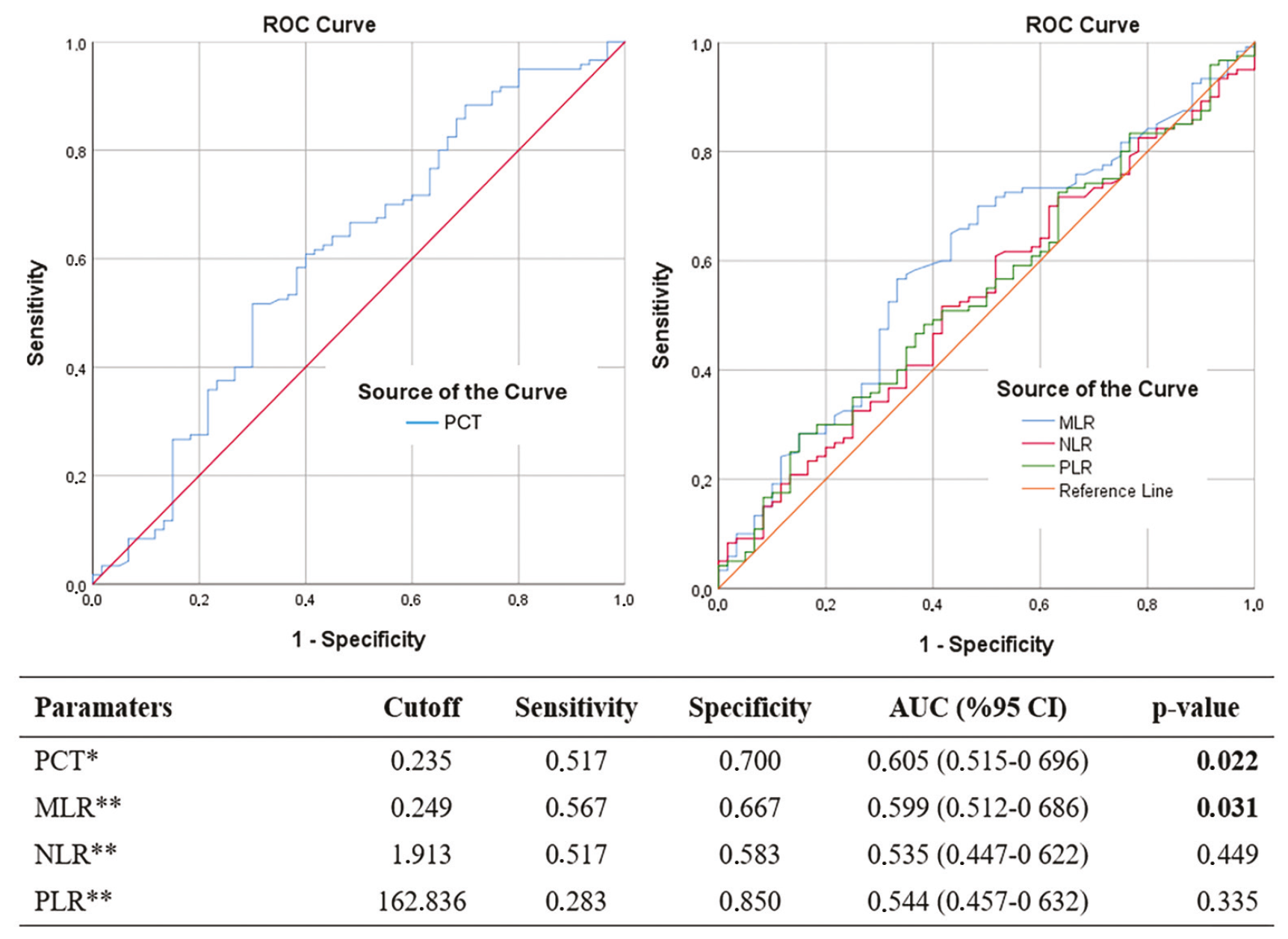
*: Smaller test values indicate a more positive test result, **: Larger test values indicate a more positive test result. AUC: Area under the curve, CI: Confidence interval, PCT: Plateletcrit, MLR: Monocyte-to-lymphocyte ratio, NLR: Neutrophil-to-lymphocyte ratio, PLR: Platelet-to-lymphocyte ratio, ROC: Receiver operating characteristic
In the ROC analysis conducted to discriminate ILD in primary SjD patient groups, the AUC for PCT was 0.490 (p=0.848) at diagnosis and 0.455 (p=0.398) at the 6-month follow-up. At diagnosis, PCT showed a sensitivity of 86.7% and a specificity of 33.3%, while at 6-months these values were 68.3% and 46.7%, respectively; neither time point showed statistical significance ().
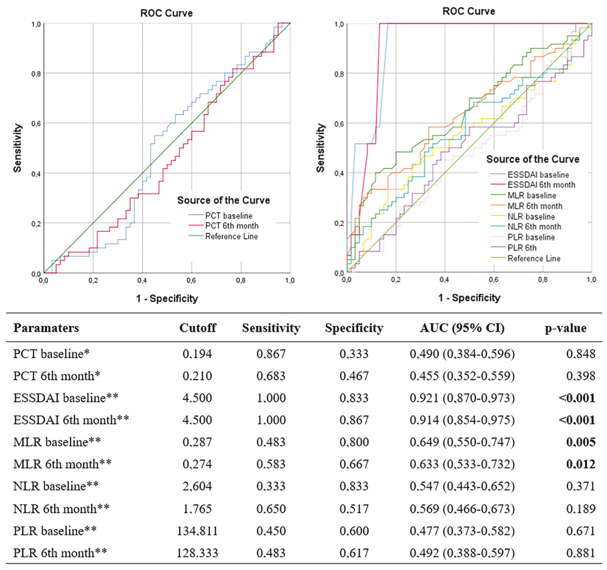
*: Smaller test values indicate a more positive test result. **Larger test values indicate a more positive test result. AUC: Area under the curve, CI: Confidence interval, ESSDAI: European League Against Rheumatism Sjögren’s syndrome disease activity index, PCT: Plateletcrit, MLR: Monocyte-to-lymphocyte ratio, NLR: Neutrophil-to-lymphocyte ratio, PLR: Platelet-to-lymphocyte ratio, ROC: Receiver operating characteristic
For MLR, the AUC values were 0.649 (p=0.003) at diagnosis and 0.633 (p=0.009) at 6-months. ESSDAI showed notably higher AUC values of 0.921 (p<0.001) at diagnosis and 0.914 (p<0.001) in 6-months. Particularly for ESSDAI, sensitivity was 100% at both diagnosis and 6-months, with specificities of 83.3% and 86.7%, respectively ().
Correlations of PCT with clinical parameters in primary SjD-ILD patients
No correlation was observed between PCT and ESSDAI at diagnosis and at the 6-month follow-up (p=0.956 and p=0.314, respectively). Likewise, at both time points, PCT showed no significant relationship with FVC (p=0.233 and p=0.351).
Discussion
In this study, lower PCT levels were observed in patients with primary Sjögren’s disease (primary SjD) compared with healthy controls. However, PCT levels did not differ significantly between primary SjD patients with ILD and those without ILD at diagnosis or at 6-months. PCT levels at diagnosis were significantly lower in the primary SjD-ILD group than in healthy controls.
Pulmonary involvement represents a key factor influencing long-term prognosis, and previous studies have reported a correlation between lung involvement and greater disease severity in primary SjD (, ). However, lung involvement in patients with primary SjD can be nonspecific, which may lead to delay in diagnosis. Since ILD is considered to be associated with early mortality, identifying risk factors for these diseases is of great importance (, ).
Consistent with a previous study (), NSIP was the most common radiological pattern identified by HRCT in our study, whereas UIP and LIP were less frequent. Abnormal PFT results were noted in primary SjD-ILD patients, as previously reported (), primarily associated with diffusion dysfunction and a restrictive pattern. However, no correlation was observed between PCT and FVC or ESSDAI. Advanced age and female sex were more frequent in primary SjD-ILD patients, in agreement with earlier findings ().
Early detection of SjD and pulmonary involvement using accessible and cost-effective tests may help prevent disease progression and contribute to better health outcomes. CBC is a cost-effective, widely available, and commonly performed laboratory test. Laboratory markers, including ESR and CRP levels, are among the most commonly used methods for evaluating disease activity in primary SjD patients (). However, these markers have certain limitations, such as reflecting only short-term inflammatory activity and having a low ability to distinguish between overlapping inflammatory conditions (). Recent studies have demonstrated that NLR, MLR, and PCT values also serve as markers of inflammation, similar to CRP and ESR (, ).
NLR and PLR values, calculated from CBC parameters, are inflammatory markers that, like MPV, PDW, and PCT, have been widely applied for diagnostic and prognostic purposes in malignancies, cardiovascular disorders, and infectious diseases (, ). Leukopenia has been reported in 12-22% of primary SjD cohorts, which is mainly mild (). In addition, neutrophils are important mediators of inflammation and have been implicated in the pathogenesis of autoimmune diseases such as RA, SLE, and primary SjD (). In a study, neutropenia (<2.5×10⁹/L) was observed in 33% of patients, with just 3% linked to neoplasia or medications, and 30% classified as idiopathic neutropenia (). In our study, although leukocyte and neutrophil counts did not differ significantly between SjD patients and the healthy control group, they were observed to be lower in patients with SjD. The triple-group analysis revealed a significant difference in leukocyte counts.
In autoimmune diseases, dense lymphocytic tissue infiltration has been observed and may contribute to decreased circulating cell counts (). Lymphocytes play a crucial role in the pathogenesis of primary SjD, being involved in local autoantibody production and linked to an increased likelihood of lymphoma (). Monocytes, as principal components of the innate immune system, bridge innate and adaptive immunity by secreting proinflammatory and proliferative cytokines and chemokines (). In autoimmune conditions, monocyte counts in peripheral blood are often reduced due to their recruitment and accumulation in affected tissues (). In this study, lymphocyte and monocyte counts were elevated in SjD patients relative to healthy controls, although the differences were not statistically significant. The observed findings may reflect the presence of ILD among study participants as well as the impact of concurrent treatments.
Parameters related to platelets, such as PLT, PCT, MPV, and PLR, are known to be linked with the activity and severity of various diseases (, ). In a study including 380 patients with primary SjD, thrombocytopenia was reported in 60 individuals (). Although platelet counts were lower in the primary SjD group, the difference was not statistically significant in our study.
In the three-group comparative analysis including SjD patients and those with ILD, MPV values were significantly reduced, in agreement with previous reports. Low MPV, similar to NLR and PLR, is considered an inflammatory indicator and is generally associated with lower disease activity in rheumatic diseases, including SLE, RA, and ankylosing spondylitis (, ). PCT, an inflammatory marker derived from CBC, is recognized to exhibit prognostic and diagnostic significance in various diseases, including gastrointestinal and cardiovascular diseases, and autoimmune disorders (, ). Increased PCT levels have been shown to be associated with disease activity in TAK (). Previous studies have shown that PCT is significantly elevated in autoimmune diseases, including RA (, ). Monitoring platelet counts in primary SjD is important for assessing systemic involvement and the need for therapeutic intervention. Autoimmune thrombocytopenia may occur in SjD, and PCT generally decreases in parallel with platelet count. However, chronic inflammation in SjD may enhance platelet activation, leading to an increase in mean platelet volume (MPV); consequently, despite a reduced platelet count, PCT may remain within or close to the normal range due to elevated MPV. We found that PCT levels were observed to be lower in primary SjD patients than in the control group. In the triple comparative analysis, PCT remained significant and was observed to be lower in both primary SjD-ILD and non-ILD patients relative to healthy controls. Although the strength of ROC analysis is weak, PCT may discriminate SjD. However, its discriminative capacity for ILD in primary SjD patients was not significant. This lack of significance may be attributed to the confounding effects of immunosuppressive treatments received by primary SjD-ILD patients.
MLR levels were significantly higher in the primary SjD group compared with healthy controls. MLR was identified as a discriminative marker for SjD (). Also, elevated hematological ratios such as NLR and MLR have been shown to exhibit positive correlations with systemic inflammation indicators and disease activity in various autoimmune diseases, including RA, systemic sclerosis, inflammatory myopathies, and primary SjD (). In addition, patients with primary SjD-ILD showed significantly higher MLR values. ROC analysis indicated that MLR outperforms other ratios in discriminating patients with primary SjD-ILD, exhibiting higher sensitivity at the established cut-off value.
The association between hematological indices and ESSDAI has not yet been explored (). In our study, ESSDAI was evaluated both at diagnosis and at the 6-month follow-up. Significant differences were observed between groups based on the presence of ILD. ESSDAI at diagnosis was correlated with leukocyte count and MLR, whereas ESSDAI at 6 months was correlated with leukocyte and neutrophil counts. Additionally, both ESSDAI measurements were correlated with FVC, but no association was found with PCT.
Study Limitations
This study has several limitations. First, the retrospective observational study design limits the ability to infer causality between PCT and primary SjD-ILD. CBC values were obtained from newly diagnosed, early-stage, treatment-naive SjD patients. However, information regarding medications used by SjD patients with concomitant ILD from initial diagnosis to ILD diagnosis was not provided. Furthermore, laboratory values assessed at the 6-month follow-up after ILD diagnosis may have been influenced by immunosuppressive and corticosteroid treatments administered during that period. It is important to consider the effects of corticosteroids on immune cell counts and on PCT, NLR, and PLR when interpreting the results. PCT may also have prognostic value; however, as our study evaluated a 6-month follow-up period, which represents a relatively limited timeframe for the development of ILD, no definitive conclusions regarding prognosis could be drawn. Additionally, our sample size was small, and the findings reflect the experience of a single center. The absence of a correlation between PCT and primary SjD-ILD could be due to the limited sample size and potential confounding effects of treatments. Well-designed prospective studies with standardized treatment protocols are required to clarify the impact of specific therapies on PCT levels and to confirm the clinical relevance of PCT in primary SjD-ILD.
Conclusion
This study demonstrated that PCT levels were significantly lower in patients with primary SjD; however, its discriminative and predictive performance was modest. PCT was not associated with ILD involvement or disease activity, suggesting that PCT alone may have limited clinical utility as a biomarker in primary SjD. There remains a need for cost-effective, reliable, and widely available biomarkers to assess disease activity in primary SjD, particularly in cases with ILD involvement. These findings may provide a basis for future prospective studies with longer follow-up periods aimed at further evaluating PCT as a potential biomarker in primary SjD.
Footnotes
Ethics
Ethics Committee Approval: The Ethics Committee of University of Health Sciences Türkiye, Gülhane Training and Research Hospital Medical Faculty approved the study protocol (approval date: 10.09.2024; no: 2024-430).
Informed Consent: Informed consent was obtained from all participants for the use of their medical records for research purposes.
Authorship Contributions
Surgical and Medical Practices: Surgical and Medical Practices: E.Ç.G., S.Y., E.T., E.B.A., B.C.A., Ö.K., M.Ç., S.Yı., Concept: E.Ç.G., S.Y., Design: E.Ç.G., S.Y., Data Collection or Processing: E.Ç.G., S.Y., Analysis or Interpretation: E.Ç.G., S.Y., E.T., E.B.A., B.C.A., Ö.K., M.Ç., S.Yı., Literature Search: E.Ç.G., S.Y., Writing: E.Ç.G., S.Y.
Conflict of Interest: No conflict of interest was declared by the authors.
Financial Disclosure: The authors declared that this study received no financial support.
References
Longhino S, Chatzis LG, Dal Pozzolo R, Peretti S, Fulvio G, La Rocca G, et al. Sjögren’s syndrome: one year in review 2023. Clin Exp Rheumatol. 2023;41(12):2343-2356.
Flament T, Bigot A, Chaigne B, Henique H, Diot E, Marchand-Adam S. Pulmonary manifestations of Sjögren’s syndrome. Eur Respir Rev. 2016;25(140):110-123.
Gao H, Zhang XW, He J, Zhang J, An Y, Sun Y, et al. Prevalence, risk factors, and prognosis of interstitial lung disease in a large cohort of Chinese primary Sjögren syndrome patients: a case-control study. Medicine (Baltimore). 2018;97(24):e11003.
Amlani B, Elsayed G, Barvalia U, Kanne JP, Meyer KC, Sandbo N, et al. Treatment of primary Sjögren’s syndrome-related interstitial lung disease: a retrospective cohort study. Sarcoidosis Vasc Diffuse Lung Dis. 2020;37(2):136-147.
Jeny F, Brillet PY, Kim YW, Freynet O, Nunes H, Valeyre D. The place of high-resolution computed tomography imaging in the investigation of interstitial lung disease. Expert Rev Respir Med. 2019;13(1):79-94.
Harifi G, Sibilia J. Pathogenic role of platelets in rheumatoid arthritis and systemic autoimmune diseases. Perspectives and therapeutic aspects. Saudi Med J. 2016;37(4):354-360.
Sun Y, Cui X, Kong X, Chen H, Wu S, Ma L, et al. The role of plateletcrit in Takayasu arteritis: a potential biomarker for disease activity and 6-month treatment response. Int J Rheum Dis. 2023;26(12):2517-2525.
Işık M, Şahin H, Hüseyin E. New platelet indices as inflammatory parameters for patients with rheumatoid arthritis. Eur J Rheumatol. 2014;1(4):144-146.
Rossaint J, Margraf A, Zarbock A. Role of platelets in leukocyte recruitment and resolution of inflammation. Front Immunol. 2018;9:2712.
Seringec Akkececi N, Yildirim Cetin G, Gogebakan H, Acipayam C. The C-reactive protein/albumin ratio and complete blood count parameters as indicators of disease activity in patients with Takayasu arteritis. Med Sci Monit. 2019;25:1401-1409.
Lee YH, Song GG. Neutrophil-to-lymphocyte ratio, mean platelet volume and platelet-to-lymphocyte ratio in Behçet’s disease and their correlation with disease activity: a meta-analysis. Int J Rheum Dis. 2018;21(12):2180-2187.
Shiboski CH, Shiboski SC, Seror R, Criswell LA, Labetoulle M, Lietman TM, et al. 2016 American College of Rheumatology/European League Against Rheumatism Classification Criteria for primary Sjögren’s syndrome: a consensus and data-driven methodology involving three international patient cohorts. Arthritis Rheumatol. 2017;69(1):35-45.
Seror R, Bowman SJ, Brito-Zeron P, Theander E, Bootsma H, Tzioufas A, et al. EULAR Sjögren’s syndrome disease activity index (ESSDAI): a user guide. RMD Open. 2015;1(1):e000022.
Raghu G, Collard HR, Egan JJ, Martinez FJ, Behr J, Brown KK, et al. An official ATS/ERS/JRS/ALAT statement: idiopathic pulmonary fibrosis: evidence-based guidelines for diagnosis and management. Am J Respir Crit Care Med. 2011;183(6):788-824.
Budak YU, Polat M, Huysal K. The use of platelet indices, plateletcrit, mean platelet volume and platelet distribution width in emergency non-traumatic abdominal surgery: a systematic review. Biochem Med (Zagreb). 2016;26(2):178-193.
Wang Y, Hou Z, Qiu M, Ye Q. Risk factors for primary Sjögren syndrome-associated interstitial lung disease. J Thorac Dis. 2018;10(4):2108-2117.
Palm O, Garen T, Berge Enger T, Jensen JL, Lund MB, Aaløkken TM, et al. Clinical pulmonary involvement in primary Sjogren’s syndrome: prevalence, quality of life and mortality--a retrospective study based on registry data. Rheumatology (Oxford). 2013;52(1):173-179.
Weng L, Chen Y, Liang T, Lin Y, Liu D, Yu C, et al. Biomarkers of interstitial lung disease associated with primary Sjögren’s syndrome. Eur J Med Res. 2022;27(1):199.
Ansar W, Ghosh S. Inflammation and inflammatory diseases, markers, and mediators: role of CRP in some inflammatory diseases. Biology of C Reactive Protein in Health and Disease. 2016;67-107.
Arigami T, Okumura H, Matsumoto M, Uchikado Y, Uenosono Y, Kita Y, et al. Analysis of the fibrinogen and neutrophil-lymphocyte ratio in esophageal squamous cell carcinoma: a promising blood marker of tumor progression and prognosis. Medicine (Baltimore). 2015;94(42):e1702.
Yingyi Z, Jiyong P, Zhigang W, Lu F. Mean platelet volume as an inflammation marker reflecting the severity of perianal abscesses. Clin Lab. 2019;65(3).
Manganelli P, Fietta P, Quaini F. Hematologic manifestations of primary Sjögren’s syndrome. Clin Exp Rheumatol. 2006;24(4):438-448.
Fu X, Liu H, Huang G, Dai SS. The emerging role of neutrophils in autoimmune-associated disorders: effector, predictor, and therapeutic targets. MedComm (2020). 2021;2(3):402-413.
Brito-Zerón P, Soria N, Muñoz S, Bové A, Akasbi M, Belenguer R, et al. Prevalence and clinical relevance of autoimmune neutropenia in patients with primary Sjögren’s syndrome. Semin Arthritis Rheum. 2009;38(5):389-395.
Yang Z, Zhang Z, Lin F, Ren Y, Liu D, Zhong R, et al. Comparisons of neutrophil-, monocyte-, eosinophil-, and basophil- lymphocyte ratios among various systemic autoimmune rheumatic diseases. APMIS. 2017;125(10):863-871.
Sène D, Ismael S, Forien M, Charlotte F, Kaci R, Cacoub P, et al. Ectopic germinal center-like structures in minor salivary gland biopsy tissue predict lymphoma occurrence in patients with primary Sjögren’s syndrome. Arthritis Rheumatol. 2018;70(9):1481-1488.
Wolf AA, Yáñez A, Barman PK, Goodridge HS. The ontogeny of monocyte subsets. Front Immunol. 2019;10:1642.
Sequí-Sabater JM, Beretta L. Defining the role of monocytes in Sjögren’s syndrome. Int J Mol Sci. 2022;23(21):12765.
Safak S, Uslu AU, Serdal K, Turker T, Soner S, Lutfi A. Association between mean platelet volume levels and inflammation in SLE patients presented with arthritis. Afr Health Sci. 2014;14(4):919-924.
Xu C, Song Z, Hu LT, Tong YH, Hu JY, Shen H. Abnormal platelet parameters in inflammatory bowel disease: a systematic review and meta-analysis. BMC Gastroenterol. 2024;24(1):214.
Gul M, Uyarel H, Akgul O, Akkaya E, Surgit O, Cakmak HA, et al. Long-term prognostic significance of admission plateletcrit values in patients with non-ST elevation myocardial infarction. Blood Coagul Fibrinolysis. 2016;27(6):696-701.
Yıldız F, Gökmen O. Haematologic indices and disease activity index in primary Sjogren’s syndrome. Int J Clin Pract. 2021;75(3):e13992.